. 2017 Jun;45(12):1594-1605.
doi: 10.1111/ejn.13602. Epub 2017 May 25.

Connexin36 localization to pinealocytes in the pineal gland of mouse and rat

Affiliations

Connexin36 localization to pinealocytes in the pineal gland of mouse and rat

S G Wang et al. Eur J Neurosci. 2017 Jun.

Abstract

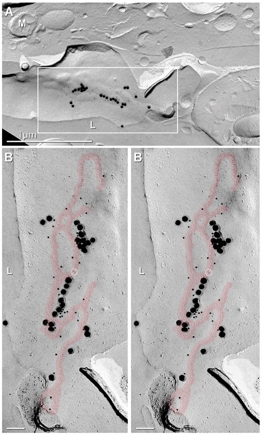
Several cell types in the pineal gland are known to establish intercellular gap junctions, but the connexin constituents of those junctions have not been fully characterized. Specifically, the expression of connexin36 (Cx36) protein and mRNA has been examined in the pineal, but the identity of cells that produce Cx36 and that form Cx36-containing gap junctions has not been determined. We used immunofluorescence and freeze fracture replica immunogold labelling (FRIL) of Cx36 to investigate the cellular and subcellular localization of Cx36 in the pineal gland of adult mouse and rat. Immunofluorescence labelling of Cx36 was visualized exclusively as puncta or short immunopositive strands that were distributed throughout the pineal, and which were absent in pineal sections from Cx36 null mice. By double immunofluorescence labelling, Cx36 was localized to tryptophan hydroxylase-positive and 5-hydroxytryptamine-positive pinealocyte cell bodies and their large initial processes, including at intersections of those processes and at sites displaying a confluence of processes. Labelling for the cell junction marker zonula occludens-1 (ZO-1) either overlapped or was closely associated with labelling for Cx36. Pinealocytes thus form Cx36-containing gap junctions that also incorporate the scaffolding protein ZO-1. FRIL revealed labelling of Cx36 at ultrastructurally defined gap junctions between pinealocytes, most of which was at gap junctions having reticular, ribbon or string configurations. The results suggest that the endocrine functions of pinealocytes and their secretion of melatonin is supported by their intercellular communication via Cx36-containing gap junctions, which may now be tested by the use of Cx36 null mice.

Keywords: gap junctions; immunofluorescence; melatonin; zonula occludens-1.


Conflict of interest statement

Competing financial interests

The authors declare no competing financial interests.

Figures

Fig. 1
Fig. 1
Immunofluorescence labelling of Cx36 in the pineal of adult mouse (C57BL/6) and rat. (A,B) Sections of mouse whole pineal double-labelled for TrypH and Cx36, showing TrypH-positive pinealocytes (A1, arrows) and, in the same field, the distribution of Cx36 in clusters of puncta (A2, arrows) in the pineal of adult wild-type mouse. Similar images of TrypH-labelled pinealocytes (B1, arrows) from a Cx36 null mouse shows, in the same field, absence of labelling for Cx36 (B2). (C) Immunofluorescence labelling of Cx36 in rat pineal using two different antibodies against Cx36, Ab39-4200 (C1) and in the same field Ab51-6300 (C2), showing complete co-localization of labelling in overlay (C3, arrows). (D) Overview of labelling for Cx36 in rat pineal, showing relative density and size variation of labelled structures. (E) Higher magnification of Cx36 immunofluorescence in mouse pineal, showing labelling to consist of clusters of fine Cx36-puncta (arrows). (F) Immunofluorescence labelling of Cx36 along the pineal stalk of adult rat. Low magnification transverse section shows dense labelling around the pineal stalk (F1, arrows) surrounding a lumen (asterisk), and higher magnification sagittal section shows patches of labelling (F2, arrows) that consist of clusters of Cx36-puncta (F2, inset).
Fig. 2
Fig. 2
Confocal double immunofluorescence labelling of TrypH and Cx36 in the pineal of adult mouse (CD1) and rat. (A,B) Overlays of labelling for TrypH and Cx36 in mouse pineal, showing punctate appearance of labelling and dense collections of Cx36-puncta (A, arrows) among clusters of pinealocytes and at a confluence of their processes (A, arrowheads), and localization of fine Cx36-puncta to pinealocyte soma (B, arrow) and along pinealocyte processes (B, arrowheads). (C–D) Overlays of labelling for TrypH and Cx36 in rat pineal, showing high concentrations of densely packed Cx36-puncta among pinealocytes processes (C, arrows), at sites of apparent close contact between pinealocyte processes (D, arrow), and unusual strand-like labelling along pinealocyte somata and their processes (E, arrows).
Fig. 3
Fig. 3
Confocal double immunofluorescence labelling of 5-HT and Cx36 in the pineal of adult rat and mouse (CD1). (A) Images of fluorescence Nissl-stained cells (pseudocolored red) (A1) and, in the same field, labelling for 5-HT (A2) in rat pineal, showing that most Nissl-positive cells are 5-HT-positive pinealocytes, as shown in overlay (A3) (arrows). Additional labelling for 5-HT is seen in sympathetic fibers (arrowheads). (B) Confocal images from rat pineal, showing 5-HT-positive pinealocyte processes (B1, arrows) and, in the same field, strand-like labelling for Cx36 localized along those processes (B2, arrows). (C–F) Overlay images of double labelling for Cx36 and 5-HT in mouse pineal, showing dense collections of Cx36-puncta (C, arrows) localized to pinealocyte initial processes (C, arrowheads), Cx36-puncta along fine processes (D, arrowheads) including points of their intersection (D, arrows), Cx36-puncta localized to pinealocyte cell bodies (E, asterisks) including points of apparent apposition (E, arrows), and pinealocyte cell bodies (F, asterisks) with a confluence of their intermingled initial processes (F, arrowheads) displaying clusters of Cx36-puncta (F, arrow).
Fig. 4
Fig. 4
Confocal double immunofluorescence labelling of Cx36 and ZO-1 in adult rat pineal. (A) Labelling of Cx36 (A1) and in the same field ZO-1 (A2), showing co-localization of labelling at both cluster of Cx36-puncta (arrowheads) and at Cx36-strands (arrows). (B) Higher magnification of a similar configuration of labelling as in (A), showing either complete co-localization of ZO-1 with Cx36 (arrow) or strand-like ZO-1-positive structures (arrowheads) that lack labelling for Cx36, but display close proximity to Cx36-strands. (C) Overlay image of double labelling for Cx36 and ZO-1, showing areas of co-localization (arrowheads), and distinct labelling for ZO-1 at long arrays of presumptive tight junctions (arrows).
Fig. 5
Fig. 5
FRIL images of pinealocyte gap junctions labelled for Cx36. (A) Low magnification showing multiple pinealocytes (purple overlay) forming gap junctions labelled for Cx36 (red overlays) at the margin of a lumen (L) within a lobule. L, lumen; M, mitochondria. (B) Higher magnification of the inscribed boxes in A, shown as stereoscopic image of a largely P-face (P) reticular gap junction (red overlay) labelled for Cx36 by 11 10-nm gold beads and five 30-nm gold beads. Arrow denotes connexon E-face pits of the apposed cell. Barred circles represent “noise” for Cx43. (C) Additional rotation and tilt of this periluminal area revealed extensive E-face (E) gap junction strands labelled for Cx36. Calibration bars are 0.1 μm unless otherwise noted.
Fig. 6
Fig. 6
FRIL images revealing E-face of Cx36-labelled pinealocyte gap junction. (A) Low magnification overview of large reticular gap junction between two pinealocytes bordering the lumen (L). M, mitochondria. (B) Higher magnification stereoscopic image of the inscribed box in A, showing an E-face gap junction (red overlay) labelled for Cx36 by 27 to 33 10-nm gold and by 14 to 26 30-nm gold beads. (For documentation of the basis for “cryptic” labelling of intact connexons remaining beneath E-face pits, see (Fujimoto, 1995; Kamasawa et al., 2006). Barred circle represents “noise” for Cx43 on the wrong side of the replica. Calibration bars are 0.1 μm unless otherwise noted.

References

    1. Bell CL, Murray SA. Adrenocortical gap junctions and their functions. Front Endocrinol. 2016;7:82. doi: 10.3389/fendo.201600082. - DOI - PMC - PubMed
    1. Belluardo N, Mudo G, Trovato-Salinaro A, le Gurun S, Charollais A, Serre-Beinier V, Amato G, Haefliger JA, Meda P, Condorelli DF. Expression of connexin36 in the adult and developing rat brain. Brain Res. 2000;865:121–138. - PubMed
    1. Berthoud VM, Saez JC. Changes in connexin43, the gap junction protein of astrocytes, during development of the rat pineal gland. J Pineal Res. 1993;14:67–72. - PubMed
    1. Berthoud VM, Hall DH, Strahsburger E, Beyer EC, Saez JC. Gap junctions in the chicken pineal gland. Brain Res. 2000;861:257–270. - PubMed
    1. Bosco D, Haefliger JA, Meda P. Connexins: key mediators of endocrine function. Physiol Rev. 2011;91:1393–1445. - PubMed

Publication types