

Molecular stretching modulates mechanosensing pathways
- PMID: 28474792
- PMCID: PMC5477536
- DOI: 10.1002/pro.3188
Molecular stretching modulates mechanosensing pathways
Abstract
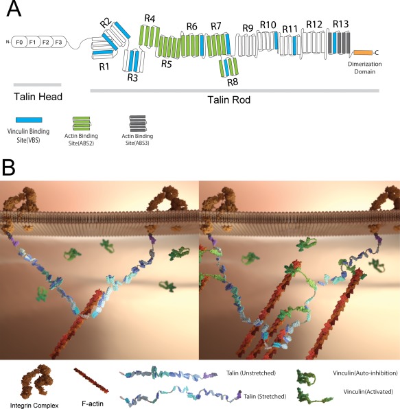
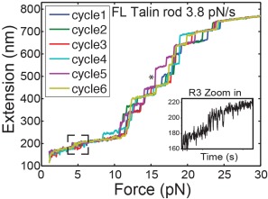
For individual cells in tissues to create the diverse forms of biological organisms, it is necessary that they must reliably sense and generate the correct forces over the correct distances and directions. There is considerable evidence that the mechanical aspects of the cellular microenvironment provide critical physical parameters to be sensed. How proteins sense forces and cellular geometry to create the correct morphology is not understood in detail but protein unfolding appears to be a major component in force and displacement sensing. Thus, the crystallographic structure of a protein domain provides only a starting point to then analyze what will be the effects of physiological forces through domain unfolding or catch-bond formation. In this review, we will discuss the recent studies of cytoskeletal and adhesion proteins that describe protein domain dynamics. Forces applied to proteins can activate or inhibit enzymes, increase or decrease protein-protein interactions, activate or inhibit protein substrates, induce catch bonds and regulate interactions with membranes or nucleic acids. Further, the dynamics of stretch-relaxation can average forces or movements to reliably regulate morphogenic movements. In the few cases where single molecule mechanics are studied under physiological conditions such as titin and talin, there are rapid cycles of stretch-relaxation that produce mechanosensing signals. Fortunately, the development of new single molecule and super-resolution imaging methods enable the analysis of single molecule mechanics in physiologically relevant conditions. Thus, we feel that stereotypical changes in cell and tissue shape involve mechanosensing that can be analyzed at the nanometer level to determine the molecular mechanisms involved.
Keywords: bioimaging; dSTORM; localization microscopy; mechanobiology; mechanoenzymatics; mechanosensing; molecular forces; protein stretching; single molecule.
© 2017 The Protein Society.
Figures
References
-
- Geiger B, Spatz JP, Bershadsky A (2009) Environmental sensing through focal adhesions. Nat Rev Mol Cell Biol 10:21–33. - PubMed
Publication types
MeSH terms
Substances
LinkOut - more resources
Full Text Sources
Other Literature Sources
