. 2017 May 5;12(5):e0177297.
doi: 10.1371/journal.pone.0177297. eCollection 2017.

Analysis of the functional compatibility of SIV capsid sequences in the context of the FIV gag precursor

Affiliations

Analysis of the functional compatibility of SIV capsid sequences in the context of the FIV gag precursor

César A Ovejero et al. PLoS One. .

Abstract

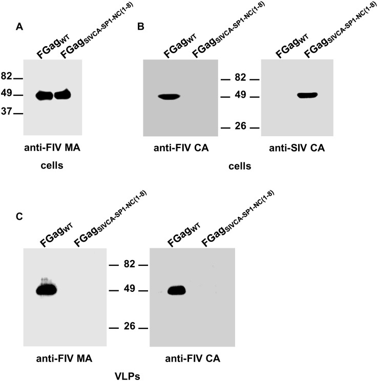
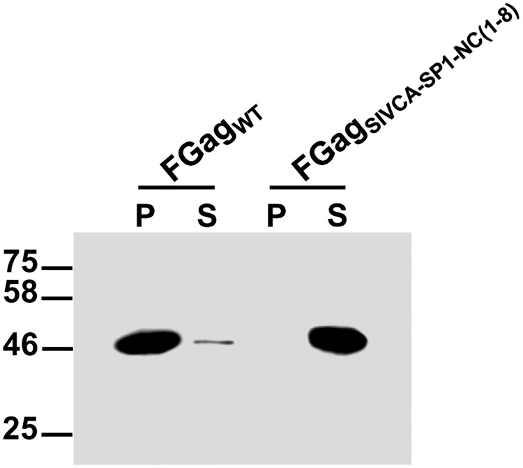
The formation of immature lentiviral particles is dependent on the multimerization of the Gag polyprotein at the plasma membrane of the infected cells. One key player in the virus assembly process is the capsid (CA) domain of Gag, which establishes the protein-protein interactions that give rise to the hexagonal lattice of Gag molecules in the immature virion. To gain a better understanding of the functional equivalence between the CA proteins of simian and feline immunodeficiency viruses (SIV and FIV, respectively), we generated a series of chimeric FIV Gag proteins in which the CA-coding region was partially or totally replaced by its SIV counterpart. All the FIV Gag chimeras were found to be assembly-defective; however, all of them are able to interact with wild-type SIV Gag and be recruited into extracellular virus-like particles, regardless of the SIV CA sequences present in the chimeric FIV Gag. The results presented here markedly contrast with our previous findings showing that chimeric SIVs carrying FIV CA-derived sequences are assembly-competent. Overall, our data support the notion that although the SIV and FIV CA proteins share 51% amino acid sequence similarity and exhibit a similar organization, i.e., an N-terminal domain joined by a flexible linker to a C-terminal domain, their functional exchange between these different lentiviruses is strictly dependent on the context of the recipient Gag precursor.


Conflict of interest statement

Competing Interests: The authors have declared that no competing interests exist.

Figures

Fig 1
Fig 1. Chimeric FIV Gag polyproteins generated to investigate the functional relationship between the FIV and SIV CA domains.
(A) Alignment of the amino acid sequences of the FIV (Petaluma isolate) and SIV (molecular clone SIVSMMPBj1.9) CA domains of Gag. The residue numbering is relative to the first amino acid (Pro) of the mature CA proteins. Identical amino acids present at the same position in both CA proteins are highlighted. Asterisks denote conservative amino acid substitutions. The open box corresponds to the MHR of the CA proteins. The NTD and CTD α-helices found in the HIV-1 CA structure [40,56] are indicated with red and blue lines, respectively, under the alignment. (B) Schematic diagram of the FIV Gag chimeras analyzed in this work. The organization of the wild-type FIV and SIV Gag precursors is depicted at the top showing the structurally conserved domains (MA, CA, and NC), the C-terminal domains (p2 in FIV Gag; p6 in SIV Gag), as well as the spacer peptides (p1 in FIV Gag; SP1 and SP2 in SIV Gag). The positions of the MHR in CA, and the N-terminal (ZFN) and C-terminal (ZFC) zinc-finger motifs in the NC domains of both FIV and SIV Gag proteins are indicated: The numbers refer to the length of each of the chimeric FIV Gag polyproteins, with residue 1 being the initiator methionine in Gag.
Fig 2
Fig 2. Effect of substituting the SIV CA-SP1-NC(1–8) region for its FIV equivalent on the production of extracellular VLPs.
COS-7 cells infected with the vTF7-3 recombinant vaccinia virus were transfected with the plasmid directing the expression of wild-type FIV Gag (FGagWT) or FIV GagSIVCA-SP1-NC(1–8) (FGagSIVCA-SP1-NC(1–8)). Thirty hours post-transfection, cells were harvested and VLPs were purified from the culture supernatants as explained in Materials and Methods. Cell (A and B) and VLP (C) lysates were analyzed for the presence of Gag proteins by Western blotting using the anti-FIV MA serum (A and C), the MAb specific for the FIV CA-CTD (B and C; indicated as anti-FIV CA), or the anti-SIV CA MAb KK60 directed to the SIV CA MHR to unambiguously detect the FIV GagSIVCA-SP1-NC(1–8) chimera (B; indicated as anti-SIV CA). Numbers indicate the positions of the molecular weight standards (in kDa). Results shown are representative of three independent experiments.
Fig 3
Fig 3. In vitro assembly reaction for recombinant FIV GagSIVCA-SP1-NC(1–8).
The purified His-FIV GagSIVCA-SP1-NC(1–8) (FGagSIVCA-SP1-NC(1–8)) and His-FIV Gagwild-type (FGagWT) proteins were incubated in parallel under the conditions described in Materials and Methods and the assembly mixtures were separated by centrifugation into the pellet (P) and supernatant (S) fractions which were then subjected to SDS-PAGE followed by Western blotting using the anti-FIV MA serum. Results shown are representative of three independent experiments.
Fig 4
Fig 4. Analysis of the association capacity of FIV GagSIVCA-SP1-NC(1–8) with wild-type FIV Gag.
COS-7 cells infected with the vTF7-3 recombinant vaccinia virus were transfected with the plasmid expressing FIV GagSIVCA-SP1-NC(1–8) (FGagSIVCA-SP1-NC(1–8)) or cotransfected with the constructs coding for wild-type FIV Gag (FGagWT) and FIV GagSIVCA-SP1-NC(1–8). As controls, cells were transfected in parallel with the plasmids encoding either wild-type FIV Gag or wild-type SIV Gag (SGagWT). Protein blots of cell (A) and VLP (B) lysates were probed with the antibodies specific for the FIV or SIV CA-CTD (indicated in the panels as anti-FIV CA and anti-SIV CA, respectively). The relative mobilities of both wild-type FIV Gag and SIV Gag as well as that of the chimeric FIV Gag protein are shown. Numbers refer to the positions of the molecular weight standards (in kDa). Results shown are representative of three independent experiments.
Fig 5
Fig 5. Ability of the chimeric FIV GagSIVCA-SP1-NC(1–8) to interact with wild-type SIV Gag in cell cultures.
COS-7 cells infected with the vTF7-3 recombinant vaccinia virus were transfected with plasmids expressing either wild-type SIV Gag (SGagWT) or FIV GagSIVCA-SP1-NC(1–8) (FGagSIVCA-SP1-NC(1–8)), or cotransfected with both plasmids. The Gag proteins in the cell (A) and VLP (B) lysates were detected by Western blotting using the anti-SIV CA MAb KK60. The relative mobilities of the wild-type and chimeric Gag proteins are shown, as are the positions of the molecular weight markers (in kDa). Results shown are representative of three independent experiments.
Fig 6
Fig 6. Comparative analysis of the effect of exchanging the FIV and SIV CA-CTD on chimeric Gag assembly.
COS-7 cells infected with the vTF7-3 recombinant vaccinia virus were transfected with plasmids directing the synthesis of wild-type FIV Gag (FGagWT) or FIV GagSIVCA(CTD) (FGagSIVCA(CTD)) (A), and, in parallel, with plasmids coding for wild-type SIV Gag (SGagWT) or SIV GagFIVCA(CTD) (SGagFIVCA(CTD)) (B). Cell and VLP lysates were analyzed for the presence of Gag proteins by Western blotting using the anti-FIV MA serum (A), or the anti-SIV MA serum and the anti-FIV CA MAb (B). Numbers indicate the positions of the molecular weight standards (in kDa). Results shown are representative of three independent experiments.
Fig 7
Fig 7. Interaction of the chimeric FIV GagSIVCA(CTD) polyprotein with wild-type SIV and FIV Gag.
COS-7 cells infected with the vTF7-3 vaccinia virus were transfected with the plasmid coding for wild-type SIV Gag (SGagWT) alone, or together with that expressing FIV GagSIVCA(CTD) (FGagSIVCA(CTD)) (A and B). In parallel, cells were transfected with the construct coding for wild-type FIV Gag (FGagWT) alone or together with the plasmid expressing FIV GagSIVCA(CTD) (C and D). Cell- and VLP-associated proteins were resolved by SDS-PAGE and the Gag proteins were visualized by Western blotting using the anti-FIV MA serum, or the MAbs targeted to the FIV or SIV CA-CTD (indicated in the panels as anti-FIV CA and anti-SIV CA, respectively). Numbers refer to the positions of the molecular weight standards (in kDa). Results shown are representative of three independent experiments.
Fig 8
Fig 8. Phenotypic characterization of the FIV GagSIVCA(NTD) chimera.
COS-7 cells infected with the vTF7-3 vaccinia virus were transfected with the plasmids expressing wild-type FIV Gag (FGagWT), or FIV GagSIVCA(NTD) (FGagSIVCA(NTD)) (A), or cotransfected with both plasmids (B). Cell- and VLP-associated proteins were detected by Western blotting using the anti-FIV MA serum, the MAb directed against the FIV CA-CTD (indicated as anti-FIV CA), or the MAb specific for the SIV CA-NTD. Numbers indicate the positions of the molecular weight standards (in kDa). Results shown are representative of three independent experiments.
Fig 9
Fig 9. Interaction of FIV GagSIVCA(NTD) with wild-type SIV Gag.
Cells infected with the vTF7-3 vaccinia virus were transfected with the plasmids expressing wild-type SIV Gag (SGagWT), FIV GagSIVCA(NTD) (FGagSIVCA(NTD)), or cotransfected with both plasmids. Cell and VLP lysates were analyzed for the presence of the wild-type SIV or chimeric Gag proteins by Western blotting using the MAbs directed to the SIV or FIV CA-CTD. Numbers indicate the positions of the molecular weight standards (in kDa). Results shown are representative of three independent experiments.

Similar articles

Cited by

References

    1. Sundquist WI, Kräusslich HG. HIV-1 assembly, budding, and maturation. Cold Spring Harb Perspect Med. 2012;2:a006924 10.1101/cshperspect.a006924 - DOI - PMC - PubMed
    1. Bell NM, Lever AM. HIV Gag polyprotein: processing and early viral particle assembly. Trends Microbiol. 2013;21:136–144. 10.1016/j.tim.2012.11.006 - DOI - PubMed
    1. Affranchino JL, González SA. Understanding the process of envelope glycoprotein incorporation into virions in simian and feline immunodeficiency viruses. Viruses. 2014;6:264–283. 10.3390/v6010264 - DOI - PMC - PubMed
    1. González SA, Affranchino JL, Gelderblom HR, Burny A. Assembly of the matrix protein of simian immunodeficiency virus into virus-like particles. Virology. 1993;194:548–556. 10.1006/viro.1993.1293 - DOI - PubMed
    1. González SA, Affranchino JL. Substitution of leucine 8 in the simian immunodeficiency virus matrix protein impairs particle formation without affecting N-myristylation of the Gag precursor. Virology. 1998; 240: 27–35. 10.1006/viro.1997.8919 - DOI - PubMed

MeSH terms

LinkOut - more resources