. 2017 Dec;13(12):1397-1409.
doi: 10.1016/j.jalz.2017.03.010. Epub 2017 May 2.

Peripheral complement interactions with amyloid β peptide: Erythrocyte clearance mechanisms

Affiliations

Peripheral complement interactions with amyloid β peptide: Erythrocyte clearance mechanisms

William D Brubaker et al. Alzheimers Dement. 2017 Dec.

Abstract

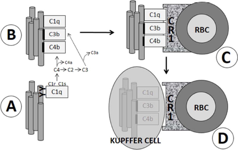
Introduction: Although amyloid β peptide (Aβ) is cleared from the brain to cerebrospinal fluid and the peripheral circulation, mechanisms for its removal from blood remain unresolved. Primates have uniquely evolved a highly effective peripheral clearance mechanism for pathogens, immune adherence, in which erythrocyte complement receptor 1 (CR1) plays a major role.

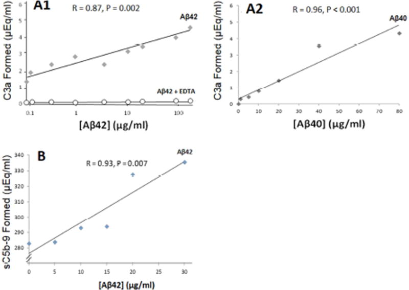
Methods: Multidisciplinary methods were used to demonstrate immune adherence capture of Aβ by erythrocytes and its deficiency in Alzheimer's disease (AD).

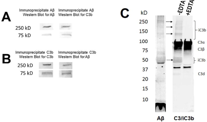
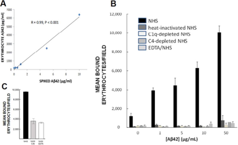
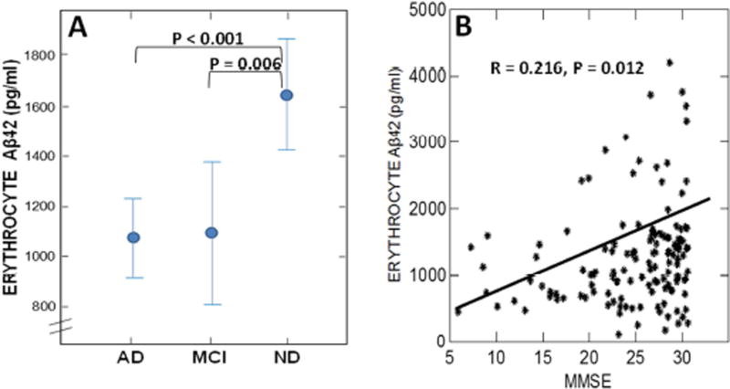
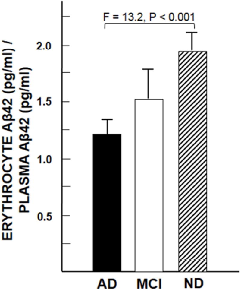
Results: Aβ was shown to be subject to immune adherence at every step in the pathway. Aβ dose-dependently activated serum complement. Complement-opsonized Aβ was captured by erythrocytes via CR1. Erythrocytes, Aβ, and hepatic Kupffer cells were colocalized in the human liver. Significant deficits in erythrocyte Aβ levels were found in AD and mild cognitive impairment patients.

Discussion: CR1 polymorphisms elevate AD risk, and >80% of human CR1 is vested in erythrocytes to subserve immune adherence. The present results suggest that this pathway is pathophysiologically relevant in AD.

Keywords: Alzheimer's disease; Amyloid β peptide; Blood; Complement; Complement receptor 1; Erythrocyte; Human; Immune adherence.


Conflict of interest statement

CONFLICTS OF INTEREST: None

Figures

Fig. 1
Fig. 1. Simplified schematic of classical pathway complement activation and immune adherence
(A) An epitope on pathogens (gray tubes) is bound by circulating antibodies (YY) specific to it. C1, the first component of the classical complement pathway, then binds to closely-apposed antibodies, forming an immune complex (IC). Notably, like certain bacterial and tumor antigens [24], Aβ has been shown to bind C1 [23] and to induce activation of the C1r and C1s proteases without antibody mediation [–23]. C1s-mediated activation of the classical complement pathway ensues, including generation of C4b, C3b, and iC3b, which become covalently fixed to the antigen (black bars). C1q also remains bound to the antigen. The antigen and/or IC is therefore said to be “opsonized” by complement (B). C) Primate (but not subprimate) erythrocytes (RBC) express cell-surface CR1, which has C4b, C3b, and C1q as ligands. Antigen/complement complexes thus become bound to erythrocytes. D) Erythrocytes then ferry the complex through the bloodstream until they reach specialized macrophages, Kupffer cells, lining the hepatic sinusoids. Kupffer cells recognize the complement tag via cell surface CRIg receptors and strip off and degrade the opsonized antigen [11,12,43].
Fig. 2
Fig. 2. Antibody-independent activation of the complement cascade
A1) Aggregated Aβ42 was incubated with NHS, then assayed by ELISA for production of C3a, a cleavage product generated from C3 following C3 activation. A significant dose-dependent response was obtained. Incubation of Aβ and serum with 10 mM EDTA, which blocks complement activation, abolished the response to Aβ and gave only background readings. A2) Aβ40 gave similar results. B) These findings were also extended to the terminal step in classical and alternative pathway activation, formation of C5b-9, the membrane attack complex, and its soluble form, sC5b-9. Significant dose-dependent activation was observed in all experiments.
Fig. 3
Fig. 3. Complement opsonization of Aβ in blood
A) In blood samples from a non-human primate inoculated with Aβ, immunoprecipitation with an anti-Aβ antibody retrieved two major bands of Aβ immunoreactivity at ~75 kD and >250 kD (left lane) and two major bands of putative C3b immunoreactivity at the same molecular weights (right lane). B) Likewise, immunoprecipitation with an anti-C3b antibody retrieved two major bands of Aβ immunoreactivity at ~75 kD and >250 kD (left lane) and two major bands of putative C3b immunoreactivity (right lane) at the same molecular weights. C) Western blot of Aβ incubated with NHS using an antibody directed against Aβ (left lane) and a Western blot of the same solution using an antibody that reacts with C3 and iC3b (right two lanes). C3 is abundantly present whether complement activation has occurred or not. In SDS/PAGE gels under reducing conditions, its two major, disulfide-linked chains, C3α and C3β, therefore dominate the gel, and, as endogenous constituents, are not affected by EDTA. By contrast, generation and covalent binding of iC3b to activating substrates such as Aβ requires complement activation and is sensitive to EDTA. Thus, putative immunoreactivity for iC3b and its fragments (brackets) is present when complement activation is permitted (−EDTA), and absent when activation is inhibited (+EDTA).
Fig. 4
Fig. 4. Erythrocyte binding and capture of Aβ
A) Following complement activation, Aβ is opsonized, tagging it for immune adherence reactions with erythrocytes. Here, NHS was incubated with various concentrations of Aβ42 to permit complement activation and opsonization, then incubated with erythrocytes from the same subject. Erythrocytes captured the Aβ42 in a significant dose-dependent manner. B) When Aβ42 was incubated with NHS, permitting complement activation and opsonization of the Aβ, significant dose-dependent binding to erythrocytes was observed (R = 0.91, P = 0.03). By contrast, binding was reduced to background with heat-inactivated NHS, which abolishes complement reactions. To control for the possibility that heat inactivation might also somehow inhibit non-complement-mediated erythrocyte capture of Aβ, we also included use of C1-depleted and C4-depleted NHS, which are specific to complement reactions, as well as EDTA treatment, a standard inhibitor of complement reactions. These conditions also abolished erythrocyte binding to Aβ, showing that complement mediation and formation of CR1 ligands is a primary mechanism for Aβ binding to erythrocytes. C) An excess of C3b, one of the ligands for CR1, was used to block erythrocyte C3b/CR1 binding sites, resulting in 62% inhibition of erythrocyte adhesion to Aβ42-coated plates.
Fig. 5
Fig. 5. Plasma and erythrocyte Aβ concentrations after infusion of Aβ40 into a non-human primate
Consistent with previous studies [8,9], plasma levels of Aβ40 spiked almost immediately after inoculation and rapidly declined over the next 15–20 minutes (top panel). Erythrocyte levels followed a nearly identical pattern and were significantly correlated with plasma levels at both doses of Aβ (bottom panel). Clearance from the erythrocyte pathway was rapid, also consistent with previous studies [19], and was capable of reducing the high dose of Aβ from nearly 10-fold normal levels to normal levels in 15–20 minutes (bottom panel).
Fig. 6
Fig. 6. Localization of Aβ42 to hepatic Kupffer cells, the final step in immune adherence
A1–A3) Confocal microscopy of a 20 µm section of AD liver showing co-localization of CD68 immunoreactivity, a marker for Kupffer cells (KC), and Aβ immunoreactivity. B) Aβ-immunostained, toluidine blue-counterstained semi-thin section of Parkinson’s disease liver, showing Aβ-immunoreactive Kupffer cells. C) Electron micrograph of ND liver, again showing typical Kupffer cell localization with apposed erythrocytes and cytoplasmic Aβ42 (arrowheads). Deletion of primary antibodies in all experiments gave uniformly negative results (not shown). We note that, like most anti-Aβ antibodies, the 4G8 antibody employed here also reacts with APP. However, the punctate, granular, intracytoplasmic labelling in these micrographs appears to be more characteristic of Aβ than its precursor, APP.
Fig. 7
Fig. 7. Pathophysiologic significance of erythrocyte capture of Aβ in AD
(A) Confirming our previous results [25], erythrocyte capture of Aβ42 is significantly deficient in both AD and MCI patients. (B) Also in concert with our previous findings [25], there was a significant correlation of erythrocyte Aβ42 levels with cognitive status (MMSE) score. Although the data clearly exhibit too much scatter to make erythrocyte Aβ a definitive prognostic for AD, they do strongly suggest that it has pathophysiologic relevance to clinical AD progression.
Fig. 8
Fig. 8. Erythrocyte capture of Aβ relative to the amount of Aβ available in the plasma compartment
Note that in each group mean erythrocyte Aβ42 levels tend to be higher than those in plasma.

Similar articles

Cited by

References

    1. Kanekiyo T, Liu CC, Shinohara M, Li J, Bu G. LRP1 in brain vascular smooth muscle cells mediates local clearance of Alzheimer’s amyloid-beta. J Neurosci. 2012;32:16458–65. - PMC - PubMed
    1. Castellano JM, Deane R, Gottesdiener AJ, Verghese PB, Stewart FR, West T, et al. Low-density lipoprotein receptor overexpression enhances the rate of brain-to-blood Abeta clearance in a mouse model of beta-amyloidosis. Proc Natl Acad Sci U S A. 2012;109:15502–7. - PMC - PubMed
    1. DeMattos RB, Bales KR, Cummins DJ, Dodart JC, Paul SM, Holtzman DM. Peripheral anti-A beta antibody alters CNS and plasma A beta clearance and decreases brain A beta burden in a mouse model of Alzheimer’s disease. Proc Natl Acad Sci U S A. 2001;98:8850–5. - PMC - PubMed
    1. Deane R, Du Yan S, Submamaryan RK, LaRue B, Jovanovic S, Hogg E, et al. RAGE mediates amyloid-beta peptide transport across the blood-brain barrier and accumulation in brain. Nat Med. 2003;9:907–13. - PubMed
    1. Do TM, Bedussi B, Chasseigneaux S, Dodacki A, Yapo C, Chacun H, et al. Oatp1a4 and an L-thyroxine-sensitive transporter mediate the mouse blood-brain barrier transport of amyloid-beta peptide. J Alzheimers Dis. 2013;36:555–61. - PubMed

MeSH terms