. 2017 May 4;66(3):384-397.e8.
doi: 10.1016/j.molcel.2017.04.012.

Structure and Dynamics of a 197 bp Nucleosome in Complex with Linker Histone H1

Affiliations

Structure and Dynamics of a 197 bp Nucleosome in Complex with Linker Histone H1

Jan Bednar et al. Mol Cell. .

Erratum in

  • Structure and Dynamics of a 197 bp Nucleosome in Complex with Linker Histone H1.
    Bednar J, Garcia-Saez I, Boopathi R, Cutter AR, Papai G, Reymer A, Syed SH, Lone IN, Tonchev O, Crucifix C, Menoni H, Papin C, Skoufias DA, Kurumizaka H, Lavery R, Hamiche A, Hayes JJ, Schultz P, Angelov D, Petosa C, Dimitrov S. Bednar J, et al. Mol Cell. 2017 Jun 1;66(5):729. doi: 10.1016/j.molcel.2017.05.018. Mol Cell. 2017. PMID: 28575663 No abstract available.

Abstract

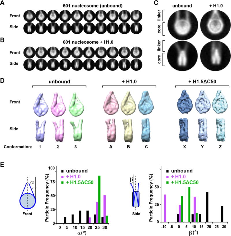
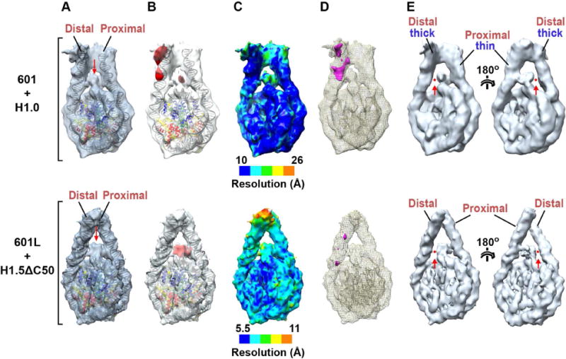
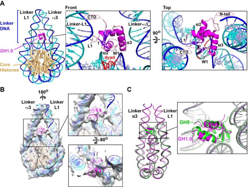
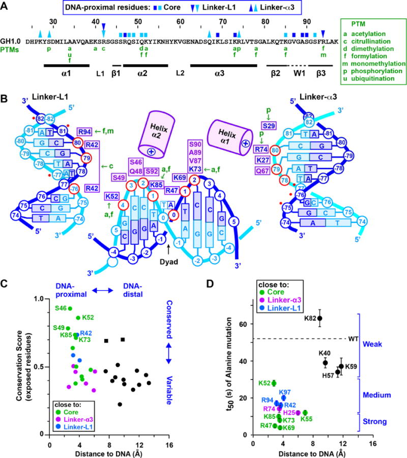
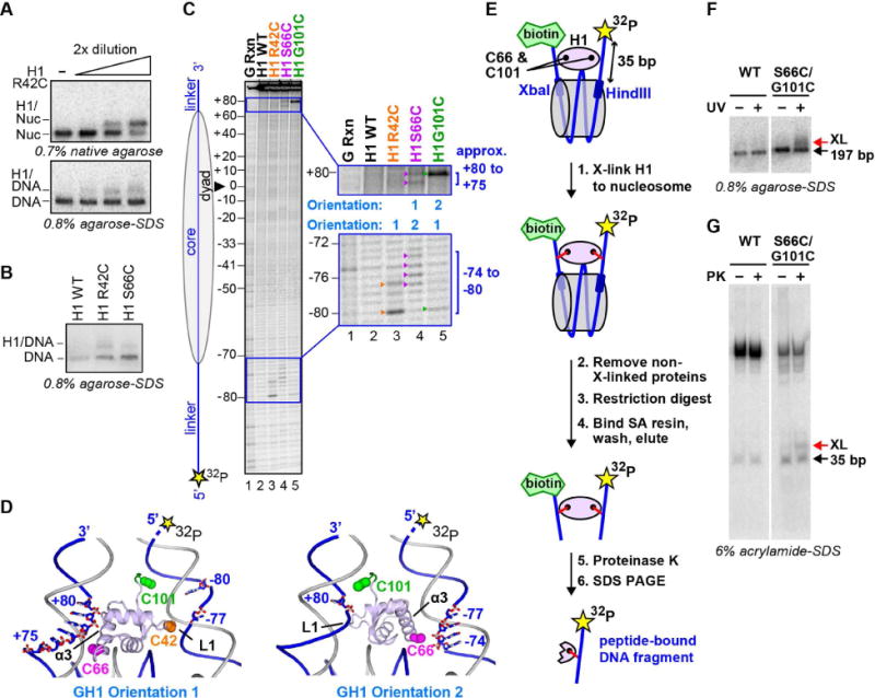
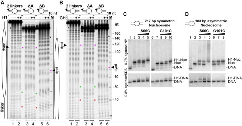
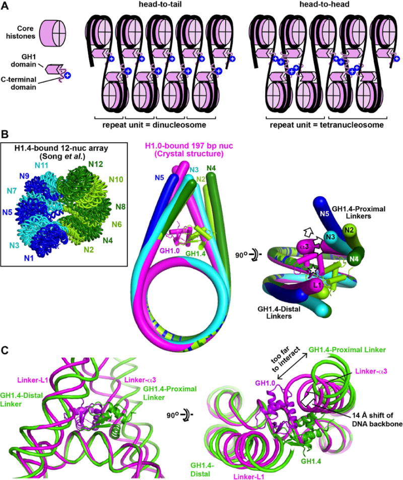
Linker histones associate with nucleosomes to promote the formation of higher-order chromatin structure, but the underlying molecular details are unclear. We investigated the structure of a 197 bp nucleosome bearing symmetric 25 bp linker DNA arms in complex with vertebrate linker histone H1. We determined electron cryo-microscopy (cryo-EM) and crystal structures of unbound and H1-bound nucleosomes and validated these structures by site-directed protein cross-linking and hydroxyl radical footprinting experiments. Histone H1 shifts the conformational landscape of the nucleosome by drawing the two linkers together and reducing their flexibility. The H1 C-terminal domain (CTD) localizes primarily to a single linker, while the H1 globular domain contacts the nucleosome dyad and both linkers, associating more closely with the CTD-distal linker. These findings reveal that H1 imparts a strong degree of asymmetry to the nucleosome, which is likely to influence the assembly and architecture of higher-order structures.

Keywords: X-ray crystallography; chromatin; cryo-EM; histone H1; hydroxyl radical footprinting; linker histone; nucleosome; protein-DNA crosslinking.


Figures

Figure 1
Figure 1. H1 stabilises a compact nucleosome conformation
(A,B) Gallery of class averages of 197-bp 601 nucleosomes in the (A) absence and (B) presence of histone H1.0. (C) Close-up views. (D) Representative 3D classes showing different linker DNA orientations in the unbound state (3 of 8 conformational classes are shown) or bound to H1.0 or H1.5ΔC50 (all 3 classes are shown). (E) Distribution of linker DNA exit angles in the unbound state (black) or bound to H1.0 (magenta) or H1.5ΔC50 (green). See also Figure S1.
Figure 2
Figure 2. Localization of H1 on the nucleosome
(A) Atomic models of the NCP and linker DNA fitted into 3D reconstructions of the H1.0-bound 601 nucleosome (top) and H1.5ΔC50-bound 601L nucleosome (bottom). (Structures are of conformations C and Y in Fig. 1D). To highlight H1-occupied density, the H1.0-bound 601 nucleosome map was bandpass filtered to keep spatial frequencies between 10 and 40 Å while that of the H1.5ΔC50-bound 601L nucleosome was sharpened by applying a negative B-factor. The red arrow indicates density attributed to the GH1 domain. (B) Density difference maps (red) calculated between the cryo-EM reconstruction and fitted atomic structures of the NCP and linker DNA. (C) Local resolution maps. (D) Difference map between the two linker arms. The proximal linker density was excised and aligned with the distal linker density. Alignment was performed at a high density threshold to favor the contribution of DNA in linker alignment. A difference map between the aligned linker arms is shown in magenta (threshold: 3 sigma). (E) Views of the H1.0-bound 601 nucleosome (top; bandpass filtered between 10 and 40 Å) and H1.5ΔC50-bound 601L nucleosome (bottom; with B-factor sharpened). Maps are displayed at a higher threshold than in panels A–D. The red arrow and dot show the loss of contact between the GH1 domain density and one of the linker arms (the thicker distal arm in the case of the 601/H1.0 complex).
Figure 3
Figure 3. Orientation of the GH1 domain
(A) Crystal structure showing the GH1.0 domain orientation. The winged-helix fold of GH1 includes a helix-turn-helix (HTH) motif formed by helices α2 and α3 and a “wing” (W1) defined by the β2-β3 loop. The base pair on the dyad axis is in red. (B) H1.0-bound 601L nucleosome crystal structure fitted into the cryo-EM map of the H1.5ΔC50-bound 601L nucleosome. (C) Alignment of the H1.0-bound nucleosome with that of chicken GH5 bound to a 167-bp nucleosome (PDB entry 4QLC)(Zhou et al., 2015). The GH1.0 and GH5 domains are related by a 10.5° rotation and by 0.5 Å shift in centre of mass. See also Figure S2 and Movies S1–S4.
Figure 4
Figure 4. Nucleosome recognition by GH1.0
(A) Primary structure of the X. laevis GH1.0 domain. Residues close to core or linker DNA are marked by blue (sense) or cyan (anti-sense) squares and triangles, respectively, coloured as in panel B. Post-translational modifications (PTMs) in mammalian histones H1.1-H1.5 (Christophorou et al., 2014; Wisniewski et al., 2007; Wisniewski et al., 2008) are in green. (B) Summary of DNA-proximal residues. GH1.0 residues are shown next to the DNA phosphate group (in red) to which they are most proximal. Residues shown are within ~4 Å of the DNA, except for Ser29 which is ~5 Å away. Basic residues are in blue, other residues in violet. The six additional linker nucleotide positions contacted by the GH5 domain in the structure of (Zhou et al., 2015) are indicated by a red dot. (C) Plot of sequence conservation versus distance from DNA for surface-exposed residues in the GH1.0 domain. For each residue, the distance from each stereochemically allowed rotamer to the closest DNA phosphate atom was measured and the shortest distance was plotted. Residues close to the core DNA or to the α3 and L1 linkers are shown in green, magenta, and blue respectively. DNA-distal residues are in black. The best-conserved residues localize close to nucleosomal DNA, while most DNA-distal residues are poorly conserved. Exceptions (conserved and DNA-distal; black squares) are Lys40, consistent with an alanine substitution of Lys40 having little effect on stability of the H1-nucleosome complex (Brown et al., 2006) (see panel D) and Ser41, which corresponds to an acidic residue in most H1 orthologs (Figure S3). (D) Effect of alanine mutations on half-time of FRAP recovery (t50) plotted versus distance from DNA. FRAP data (mean ± S.D.) are those of (Brown et al., 2006). Brackets indicate mutations with a strong, medium or weak effect on t50. See also Figure S3.
Figure 5
Figure 5. Mapping of H1-nucleosomal DNA interactions
(A to D) Site-specific cross-linking of GH1 to nucleosomal DNA. (A) Top. Native gel showing the binding of APB-derivatized H1 mutant R42C (R42C-APB) to the nucleosome. Bottom. Denaturing gel showing cross-linking of H1 R42C-APB to nucleosomal DNA after UV irradiation. (B) Denaturing gel showing cross-linking of H1 R42C-APB and H1 S66C-APB to nucleosomal DNA upon UV irradiation. (C) Mapping of cross-linked nucleotides by piperidine base elimination cleavage of the DNA and subsequent sequencing gel analysis. Nucleotides cross-linked to R42C-APB, S66C-APB and G101C-APB are indicated by orange, magenta and green arrowheads, respectively. (D) Crystal structure (orientation 1) and dyad-related orientation of GH1 (orientation 2) showing the proximity of GH1 residues to specific linker nucleotides on the radiolabeled strand. Residues 98–101 (green; absent from the crystal structure) were modeled in an extended conformation. (E to G) Simultaneous cross-linking of H1 residues to both DNA linkers. (E) Summary of the cross-linking experiment. Nucleosomes were reconstituted using 5′ biotinylated and 5′ radiolabeled 197-bp DNA containing a specific restriction endonuclease (Xba I and Hind III) site next to each linker arm. (F) APB-derivatized H1 S66C/G101C mutant binds and cross-links in a UV-dependent manner to the 197-bp nucleosome. (G) Elutions with or without proteinase K (PK) were analyzed on 6% acrylamide-SDS gel revealing a distinct band (XL) consistent with double cross-link dependent retention of the radiolabeled linker arm.
Fig. 6
Fig. 6. DNA footprinting and cross-linking analysis of H1 binding to symmetric and asymmetric nucleosomes
(A,B) Hydroxyl radical footprinting of centrally positioned nucleosomes bearing two linkers (lanes 1–2) compared to nucleosomes with only one linker (lanes 3–6). Reactions were performed in the absence (lanes 1, 3 and 5) or presence (lanes 2, 4 and 6) of (A) H1 or (B) the isolated GH1 domain. Cleavage patterns are shown in duplicate. Nucleotide regions protected by H1 or GH1 are indicated by asterisks as described in the text. (C,D) APB-derivatized H1 binding and cross-linking to (C) symmetric, 2-linker nucleosomes or (D) asymmetric, single-linker nucleosomes. Top. Native gels showing the binding of H1 S66C-APB and H1 G101C-APB to both (C) symmetric and (D) asymmetric nucleosomes. Bottom. Denaturing gels showing cross-linking of H1 S66C-APB and G101C-APB to nucleosomal DNA following UV irradiation. See also Figures S4 and S5.
Figure 7
Figure 7. Implications for higher-order chromatin structures
(A) The asymmetric localization of the CTD may influence the assembly and properties of higher-order structures. Two hypothetical arrangements shown for H1-bound nucleosomes within a two-start helical array give rise to distinct mass and electrostatic charge distributions. (B) Comparison of linker arm geometry with that observed in the condensed 12-nucleosome array of (Song et al., 2014). Nucleosomes N2-N5 of the 12-nucleosome array were aligned onto the H1.0-bound 601L nucleosome crystal structure (complex A) by superimposing the nucleosomal cores. The DNA from the crystal structure is in magenta, while that for N2-N5 are in lime, cyan, dark green and blue, respectively. (Only 4 nucleosomes of the array are shown because the 3 tetranucleosomal units have similar conformations. N5 is shown instead of N1 because the latter lacks the first linker arm). The GH1.0 domain from the crystal structure and the GH1.4 domain bound to N2 are also shown. The asterisk indicates the pseudodyad axis. The arrows show the displacement of GH1.4-proximal linkers relative to Linker-α3 of our crystal structure. The mean displacement of the DNA backbone measured one helical turn from NCP exit is 14.5 ± 6.3 Å between the GH1.4-proximal linkers and Linker-α3, and 4.0 ± 1.6 Å between the GH1.4-distal linkers and Linker-L1. (C) Comparison of linker arm geometry between the H1.0-bound crystal structure and nucleosome N2 of the 12-nucleosome array. See also Figure S6.

References

    1. Abrishami V, Vargas J, Li X, Cheng Y, Marabini R, Sorzano CO, Carazo JM. Alignment of direct detection device micrographs using a robust Optical Flow approach. J Struct Biol. 2015;189:163–176. - PubMed
    1. Adams PD, Afonine PV, Bunkoczi G, Chen VB, Davis IW, Echols N, Headd JJ, Hung LW, Kapral GJ, Grosse-Kunstleve RW, et al. PHENIX: a comprehensive Python-based system for macromolecular structure solution. Acta crystallographica. Section D. Biological crystallography. 2010;66:213–221. - PMC - PubMed
    1. Allan J, Hartman PG, Crane-Robinson C, Aviles FX. The structure of histone H1 and its location in chromatin. Nature. 1980;288:675–679. - PubMed
    1. Allan J, Mitchell T, Harborne N, Bohm L, Crane-Robinson C. Roles of H1 domains in determining higher order chromatin structure and H1 location. J Mol Biol. 1986;187:591–601. - PubMed
    1. Bednar J, Horowitz RA, Dubochet J, Woodcock CL. Chromatin conformation and salt-induced compaction: three-dimensional structural information from cryoelectron microscopy. J Cell Biol. 1995;131:1365–1376. - PMC - PubMed

MeSH terms