. 2017 May 4;169(4):597-609.e11.
doi: 10.1016/j.cell.2017.04.024.

Recurrent Potent Human Neutralizing Antibodies to Zika Virus in Brazil and Mexico

Affiliations

Recurrent Potent Human Neutralizing Antibodies to Zika Virus in Brazil and Mexico

Davide F Robbiani et al. Cell. .

Abstract

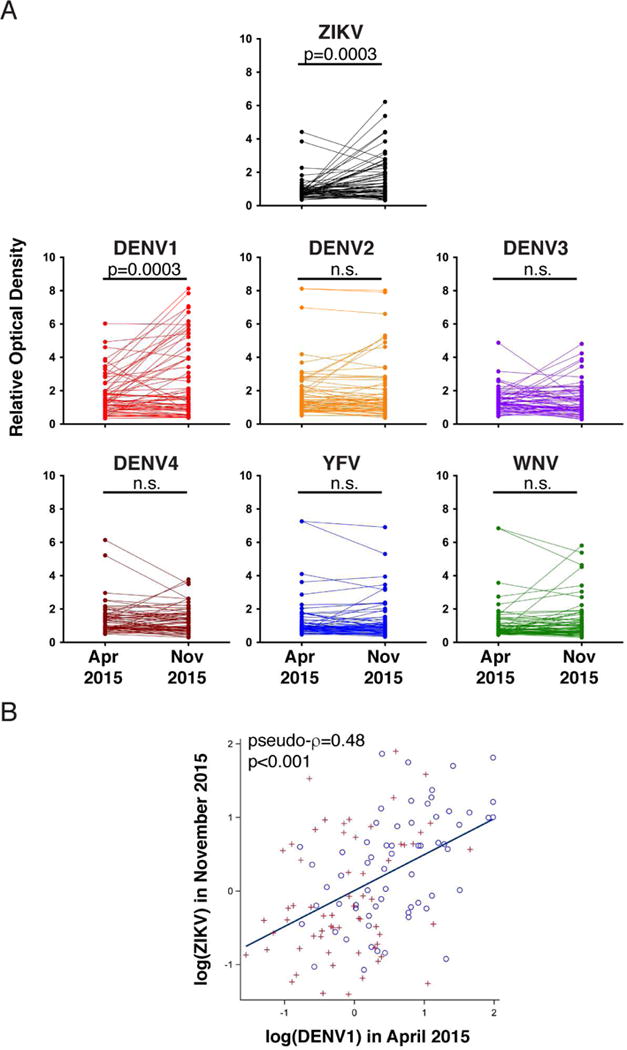
Antibodies to Zika virus (ZIKV) can be protective. To examine the antibody response in individuals who develop high titers of anti-ZIKV antibodies, we screened cohorts in Brazil and Mexico for ZIKV envelope domain III (ZEDIII) binding and neutralization. We find that serologic reactivity to dengue 1 virus (DENV1) EDIII before ZIKV exposure is associated with increased ZIKV neutralizing titers after exposure. Antibody cloning shows that donors with high ZIKV neutralizing antibody titers have expanded clones of memory B cells that express the same immunoglobulin VH3-23/VK1-5 genes. These recurring antibodies cross-react with DENV1, but not other flaviviruses, neutralize both DENV1 and ZIKV, and protect mice against ZIKV challenge. Structural analyses reveal the mechanism of recognition of the ZEDIII lateral ridge by VH3-23/VK1-5 antibodies. Serologic testing shows that antibodies to this region correlate with serum neutralizing activity to ZIKV. Thus, high neutralizing responses to ZIKV are associated with pre-existing reactivity to DENV1 in humans.

Keywords: Zika virus; antibodies; dengue virus; flavivirus; structure; vaccine.


Figures

Figure 1
Figure 1. Identification of individuals with high ZEDIII binding and neutralization capacity
A- Sera from the Brazilian and Mexican cohorts were screened by ELISA for IgG antibodies binding to ZEDIII. Each dot represents an individual donor. Optical densities are normalized to control serum from a flavivirus naïve individual vaccinated for YFV. In blue are sera selected for neutralization analysis. B- The neutralization capacity of selected sera from Mexico (red), Brazil (light red) and control samples obtained in Brazil in 2010 (grey) was determined by a ZIKV luciferase reporter viral particle (RVP) neutralization assay. The reciprocal of the serum dilution that resulted in 50% inhibition compared to RVP alone is reported as the 50% neutralization titer (NT50). The dotted line indicates the lower limit of dilutions that were examined. The five samples below the dotted line have NT50 values lower than 103. Individuals from whom antibodies were sequenced and cloned are indicated.
Figure 2
Figure 2. Discovery of ZEDIII-specific antibodies
A- Frequency of ZEDIII-specific, IgG+ memory B cells in peripheral blood of 6 donors. Flow cytometry plots display the percentage of all IgG+ memory B cells that bind to a fluorescently tagged ZEDIII bait. Flavivirus naïve peripheral blood samples are shown alongside as negative controls. B- Pie charts show the distribution of antibody clones that share the same IGHV and IGLV; the width of each colored or shaded slice is proportional to the number of clones sharing a distinct combination of IGHV and IGLV sequences. The total number of antibody clones sequenced from each donor is indicated in the center of the pie chart. VH3-23/VK1-5 clones are in red, while other VH3-23 clones are indicated with different shades of blue. Non VH3-23 clones are shown in shades of grey, and singlets are in white. None of the grey clones are recurrent across individuals. C- V(D)J assignments for the VH3-23/VK1-5 clones. IgBLAST was used to assign the germline (GL) reference sequence for IGHV and IGLV. Red highlights differences in D and J usage in the VH3-23 clones between individuals.
Figure 3
Figure 3. Binding of cloned antibodies to EDIII from ZIKV and other flaviviruses
A- Binding of human monoclonal antibodies to ZEDIII. Human anti-HIV antibody 10–1074 was used as a negative control (Mouquet et al., 2012). The average half effective concentration (EC50) from at least two independent experiments is shown. B- Somatic mutations are required for ZEDIII binding. Binding of Z004, its predicted germline (GL), and control antibodies to ZEDIII as assessed by ELISA is shown. C- Human monoclonal antibody cross-reactivity by ELISA. Reactivity to the EDIII of the indicated flaviviruses is shown in blue. The list of antibodies is reported on the left of panel A. D- Z004 binds to the EDIII of DENV1. Binding of Z004, its predicted germline (GL), and control antibodies to DENV1 EDIII as assessed by ELISA is shown.
Figure 4
Figure 4. VH3-23/VK1-5 antibodies neutralize ZIKV and DENV1
A- Neutralization potency of human monoclonal antibodies by ZIKV luciferase RVP assay. The human anti-HIV antibody 10–1074 serves as a negative control. Average values of the half maximal inhibitory concentration (IC50) from at least two independent experiments are shown. B and C- Z004 neutralizes ZIKV (B) and DENV1 (C) RVPs. Luciferase activity relative to the no antibody control was determined in the presence of increasing concentrations of Z004 or of its predicted germline antibody as indicated. Control antibody was tested at a single concentration. Data are represented as mean ± SD. D, E, and F- Z004 protects IFNAR−/− mice from ZIKV disease. Mice were infected by footpad (f.p.) injection with the Puerto Rican PRVABC59 ZIKV strain and treated intraperitoneally (i.p.) with Z004 (or 10–1074 control) either before (E) or 1 day after (F) infection. Mice were monitored for symptoms and survival. Survival: p<0.0001 (pre-exposure) and p=0.0027 (post-exposure). Symptoms: p<0.0001 (both pre- and post-exposure, Mantel-Cox test). Three independent experiments, of 4 to 7 mice per group, were combined and displayed. G- Amino acid alignment of a portion of the EDIII lateral ridge region for a panel of flaviviruses. The corresponding accession numbers are indicated in parenthesis. H- The K394 residue in the ZEDIII lateral ridge is required for ZIKV neutralization by Z004. Luciferase activity relative to no antibody control was determined for ZIKV wild type or mutant E393A and K394A RVPs. I- Z004 neutralizes both Asian/American and African strains. RVPs bearing Asian/American ZIKV wild type (E393), mutant (Asian/American with E393D) and African strain (D393) E proteins were neutralized by Z004. In H and I data are represented as mean ± SD.
Figure 5
Figure 5. Structures of Fab complexes with ZIKV and DENV1 EDIII domains
A- Superimposition of Z006 Fab-ZEDIII and Z004 Fab-DENV1 EDIII crystal structures after alignment of the EDIII domains. The VH domain positions differ by a 14° rotation about an axis passing through the center of the interface. Inset: close-up of interactions between the E393ZIKV–K394ZIKV/E384DENV1–K385DENV1 motif (shown as sticks) within the EDIII lateral ridge and the two Fabs. Fab CDRs are highlighted. B- Overlay of the Z006-ZEDIII complex structure (VH-VL in blue and cyan; EDIII in black) with previously solved structures of antibodies in complex with ZIKV and DENV1 EDIII domains. VH-VL domains from ZIKV antibodies are pink; VH-VL from DENV1 antibodies are tan; the E393ZIKV–K394ZIKV side chains in ZEDIII are shown as spheres. Structures were aligned on the EDIII domains; only ZEDIII is shown for clarity. C- ZEDIII epitope: EDIII residues contacted by Z006 Fab are highlighted on a surface representation of the EDIII structure. EDIII residues contacted by VH are blue, residues contacted by VL are cyan, and residues making interactions with both VH and VL are dark blue. The E393ZIKV–K394ZIKV motif is outlined. Contacts between the Z004 Fab and DENV1 EDIII were less extensive than Z006–ZEDIII contacts, in part because of disorder of the CC’ loop in DENV1 EDIII (residues 343–349) (Methods). D to F- Comparison of key antibody-antigen interactions for Z006 Fab-ZEDIII and Z004 Fab-DENV1 EDIII structures. Hydrogen bonds are shown as dotted lines. D- Fab interactions with K394ZIKV/K385DENV1. E- Fab interactions with E393ZIKV/E384DENV1. F- Y58HC (germline-encoded in VH3-23) interactions with antigen. See also Tables S5 and S6.
Figure 6
Figure 6. EDIII reactivity over time
A- A set (n=63) of paired sera from the Brazilian cohort participants were collected in April and November 2015 and assayed for binding to flavivirus EDIII. Optical densities are normalized as described in Fig. 1A. Paired sera from the same individual are connected by a line. Each value represents the average of two independent measurements. P values were determined with the two-tailed paired t test (n.s., not significant). B- Correlation between DENV1 EDIII reactivity in April and ZEDIII reactivity in November 2015. Circles and plus signs distinguish data from two independent experiments. Pseudo-ρ=0.48, p<0.001 by univariate analysis (see statistical methods).
Figure 7
Figure 7. EDIII antibodies contribute to the serologic response and ZIKV neutralization capacity
A- Competition ELISA shows the increase within individuals of serum antibodies that block biotin-Z004 ZEDIII binding after ZIKV exposure. Each dot represents a serum sample (n=62 at each of the indicated time points). A line connects sera from the same individual obtained at different time points. The p value was determined with the two-tailed paired t test. B and C- The estimated quantity (μg/ml) of Z004 blocking antibodies in the serum obtained after ZIKV introduction (X axes) was plotted with the overall serum binding activity to ZEDIII (Y axis, B), and the change in that individual’s serum ZEDIII binding from before to after ZIKV (Y axis, C). Binding activity change was determined by subtracting the pre- from the post-ZIKV ELISA relative optical density value (average of two independent measurements). Each dot represents an individual (n=62, two-tailed Spearman r test). D and E- Serum neutralization potency expressed as NT50 versus the overall serum binding activity to ZEDIII (D), or Z004 blocking antibody concentrations in sera (E) obtained after ZIKV introduction are plotted. Each dot represents a serum sample from a single donor (n=27, two-tailed Spearman r test). Representative of two independent experiments is shown.

Similar articles

Cited by

References

    1. Adams PD, Afonine PV, Bunkoczi G, Chen VB, Davis IW, Echols N, Headd JJ, Hung LW, Kapral GJ, Grosse-Kunstleve RW, et al. PHENIX: a comprehensive Python-based system for macromolecular structure solution. Acta crystallographica Section D, Biological crystallography. 2010;66:213–221. - PMC - PubMed
    1. Arnaout R, Lee W, Cahill P, Honan T, Sparrow T, Weiand M, Nusbaum C, Rajewsky K, Koralov SB. High-resolution description of antibody heavy-chain repertoires in humans. PloS one. 2011;6:e22365. - PMC - PubMed
    1. Barba-Spaeth G, Dejnirattisai W, Rouvinski A, Vaney MC, Medits I, Sharma A, Simon-Loriere E, Sakuntabhai A, Cao-Lormeau VM, Haouz A, et al. Structural basis of potent Zika-dengue virus antibody cross-neutralization. Nature. 2016;536:48–53. - PubMed
    1. Bardina SV, Bunduc P, Tripathi S, Duehr J, Frere JJ, Brown JA, Nachbagauer R, Foster GA, Krysztof D, Tortorella D, et al. Enhancement of Zika virus pathogenesis by preexisting antiflavivirus immunity. Science 2017 - PMC - PubMed
    1. Barzon L, Pacenti M, Franchin E, Lavezzo E, Trevisan M, Sgarabotto D, Palu G. Infection dynamics in a traveller with persistent shedding of Zika virus RNA in semen for six months after returning from Haiti to Italy, January 2016. Euro surveillance : bulletin Europeen sur les maladies transmissibles = European communicable disease bulletin 21 2016 - PMC - PubMed