. 2017 May 4;169(4):722-735.e9.
doi: 10.1016/j.cell.2017.04.020.

Molecular Mechanism of Substrate Processing by the Cdc48 ATPase Complex

Affiliations

Molecular Mechanism of Substrate Processing by the Cdc48 ATPase Complex

Nicholas O Bodnar et al. Cell. .

Abstract

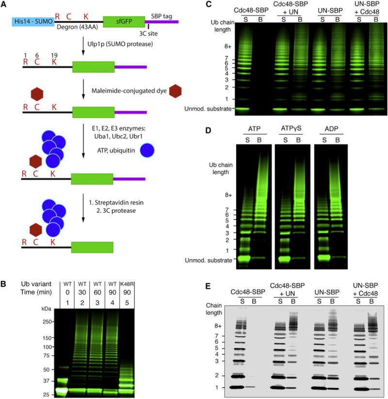
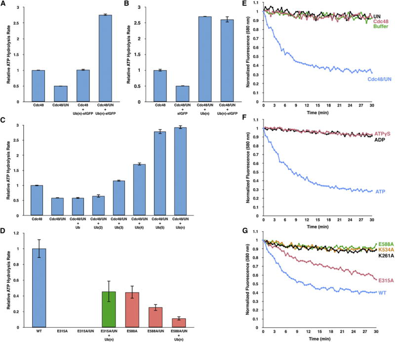
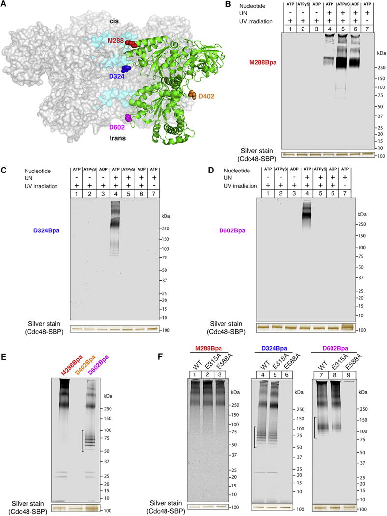
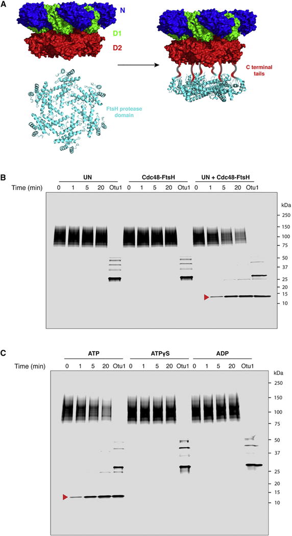
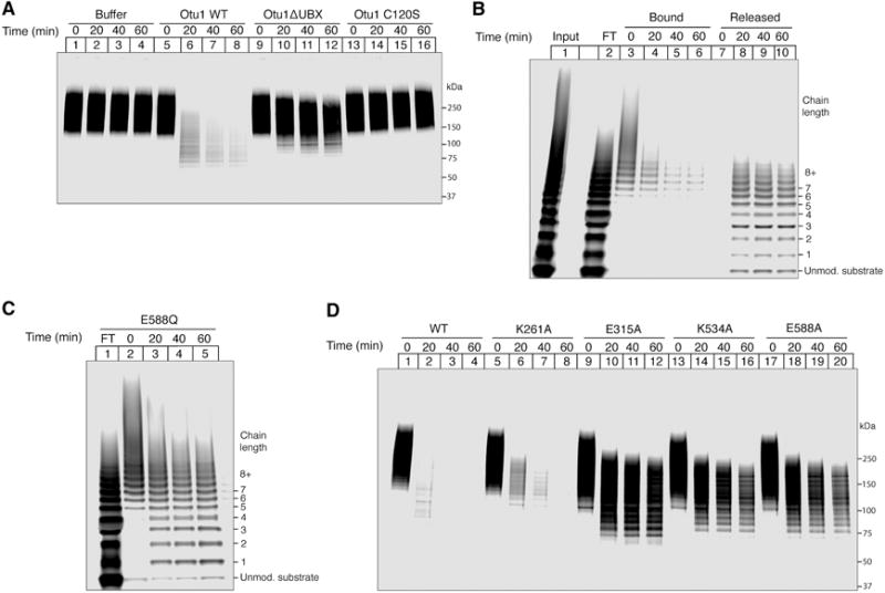
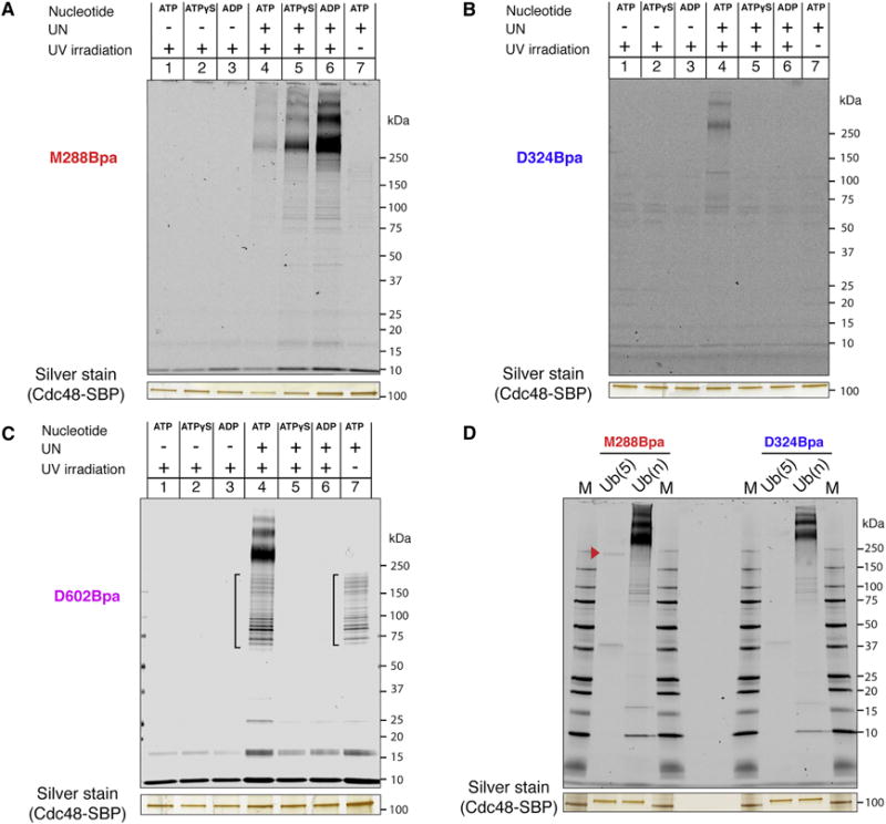
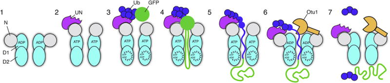
The Cdc48 ATPase and its cofactors Ufd1/Npl4 (UN) extract polyubiquitinated proteins from membranes or macromolecular complexes, but how they perform these functions is unclear. Cdc48 consists of an N-terminal domain that binds UN and two stacked hexameric ATPase rings (D1 and D2) surrounding a central pore. Here, we use purified components to elucidate how the Cdc48 complex processes substrates. After interaction of the polyubiquitin chain with UN, ATP hydrolysis by the D2 ring moves the polypeptide completely through the double ring, generating a pulling force on the substrate and causing its unfolding. ATP hydrolysis by the D1 ring is important for subsequent substrate release from the Cdc48 complex. This release requires cooperation of Cdc48 with a deubiquitinase, which trims polyubiquitin to an oligoubiquitin chain that is then also translocated through the pore. Together, these results lead to a new paradigm for the function of Cdc48 and its mammalian ortholog p97/VCP.

Keywords: AAA ATPase; ERAD; VCP; p97; proteasome; translocation; ubiuitination.


Figures

Figure 1
Figure 1. Cdc48/UN complex interacts with a polyubiquitinated substrate
(A) Scheme for the synthesis and purification of polyubiquitinated superfolder GFP (sfGFP) a Cdc48 substrate containing an N-end degron; see text for details. (B) Dye-labeled sfGFP bearing the N-end degron was incubated with ubiquitination machinery, ATP, and the indicated ubiquitin variant. Samples were analyzed by SDS-PAGE and fluorescence scanning. The unmodified substrate appears as two bands due to incomplete denaturation in SDS. (C) The indicated proteins were immobilized through a SBP tag on streptavidin beads and incubated with ubiquitinated sfGFP. Supernatants (S) and bound material (B) were analyzed by SDS-PAGE and fluorescence scanning. (D) As in (C), with Cdc48 and SBP-tagged UN used in each condition. The indicated nucleotide was included in all steps following purification. Irrelevant lanes of the gel have been removed for display (white spaces). (E) As in (C), but with free fluorescently labeled K48-linked polyubiquitin chains instead of polyubiquitinated sfGFP. See also Figure S1.
Figure 2
Figure 2. Ubiquitin stimulates Cdc48 ATPase activity for substrate unfolding
(A) ATP hydrolysis rates were determined with the indicated combinations of purified proteins and substrate (Cdc48; UN; Ub(n)-GFP). Substrate was included at 5-fold excess over the cofactor to maximize occupancy. The rates were normalized with respect to that of Cdc48 alone. Shown are the means and standard deviations of three experiments. (B) As in (A), but also with free K48-linked polyubiquitin chains (Ub(n)), carrying up to ~15 ubiquitin moieties. (C) ATPase stimulation with free K48-linked ubiquitin chains of increasing length. (D) As in (B), but with Cdc48 Walker B mutations in D1 (E315A; green bars) or D2 (E588A; red bars). E315A and E588A can bind, but not hydrolyze, ATP in D1 and D2, respectively (Walker B mutations). (E) Irradiated, fluorescent Eos, consisting of two polypeptide segments, was polyubiquitinated, as described for sfGFP (Figure 1A). This substrate (Ub(n)-Eos) was incubated with an ATP-regenerating system and excess of the indicated proteins. After addition of ATP, Eos fluorescence was followed over time. (F) As in (E), but with Cdc48 and UN present in all conditions, and the nucleotide varied as indicated. (G) As in (F), but with UN and an ATP regenerating system present in all conditions. Cdc48 or its point mutants were included as indicated. K261A and K534A are defective in ATP binding to the D1 and D2 ATPases, respectively (Walker A mutations), and E315A and E588A can bind, but not hydrolyze, ATP in D1 and D2, respectively (Walker B mutations). See also Figure S2.
Figure 3
Figure 3. Substrate passes through the central pore of Cdc48
(A) Cdc48 positions used for introduction of the Bpa crosslinker. Sections of the central pore are highlighted in cyan, and a single Cdc48 monomer in the hexamer is shown in green. The Cdc48 model was generated based on PDB ID 3CF1. (B) SBP-tagged Cdc48 (Cdc48-SBP) with Bpa at the M288 position (D1 pore loop) was incubated with dye-labeled, polyubiquitinated sfGFP and the indicated combinations of UN and nucleotide. After irradiation and pulldown with streptavidin beads, crosslinked species were detected by SDS-PAGE and fluorescence scanning. The silver-stained band of Cdc48-SBP serves as a loading control. (C) As in (B), but with Bpa at the D324 position in the interior of the central pore. (D) As in (B), but with Bpa at the D602 position in the D2 pore-2 loop. (E) As in (B), but with Bpa at an external position (D402). For comparison, crosslinking was also performed with probes in the pore. UN and ATP were included in all conditions. It should be noted that some non-crosslinked substrate remained associated with Cdc48. We generally observed that in the presence of ATP, non-crosslinked material had a tendency to stick to Cdc48 (brackets; see also Figure 3F), likely because it was present in an unfolded conformation inside the pore. (F) As in (B), but with Cdc48 mutants that contain the crosslinker and Walker mutations at the indicated positions. E315A, Walker B mutation in D1; E588A, Walker B mutation in D2. UN and ATP were included in all conditions. See also Figure S3.
Figure 4
Figure 4. Substrate exits from the D2 side of the double ring ATPase
(A) The proteolytic domain of FtsH (PDB ID 2DI4) was fused to the C-terminal tail of the Cdc48 ATPase to produce a hybrid AAA protease (Cdc48-FtsH). (B) Dye-labeled, polyubiquitinated Eos was incubated with an ATP regenerating system and the indicated proteins. Aliquots were removed at the indicated time points and analyzed by SDS-PAGE and fluorescence scanning. After the time course, a 50-fold excess of Otu1 was added to remove ubiquitin chains. The red arrow head indicates a stable fragment containing the fluorescent dye, which was generated when both Cdc48-FtsH and UN are present. The dye is attached to C238, near the C-terminus of the 275-residue substrate. (C) As in (B), with Cdc48-FtsH and UN in all conditions, and the nucleotide varied as indicated. See also Figure S4.
Figure 5
Figure 5. Substrate release from the Cdc48 complex requires deubiquitination
(A) Complexes of Cdc48, SBP-tagged UN, and dye-labeled, polyubiquitinated sfGFP were immobilized on streptavidin beads in the presence of ATP. After washing, the indicated Otu1 variants were added and samples of the bound material were analyzed at the indicated time points by SDS-PAGE and fluorescence scanning. WT, wild type; Otu1ΔUBX, Otu1 lacking the UBX domain; Otu1 C120S, catalytically inactive Otu1. (B) As in (A), but with both bound and released material analyzed over time. Lane 1 shows the input material before incubation with streptavidin beads, and lane 2 the fraction that did not bind (flow-through; FT). (C) As in (A), but with wild type Otu1 and the Walker B mutant E588Q in D2, which slowly hydrolyzes ATP. (D) As in (C), but with other Walker mutations in D1 or D2. K261A, Walker A mutation in D1; E315A, Walker B mutation in D1; K534A, Walker A mutation in D2; E588A, Walker B mutation in D2. See also Figure S5.
Figure 6
Figure 6. Ubiquitin passes through the central pore of Cdc48
(A) SBP-tagged Cdc48 (Cdc48-SBP) with Bpa at the M288 position (D1 pore loop) was incubated with dye-labeled free, K48-linked polyubiquitin chains and the indicated combinations of UN and nucleotide. After irradiation and streptavidin pulldown, crosslinked species were detected by SDS-PAGE and fluorescence scanning. The silver-stained band of Cdc48-SBP serves as a loading control. (B) As in (A), but with Bpa at the D324 position in the interior of the central pore. (C) As in (A), but with Bpa at the D602 position in the D2 pore-2 loop. Brackets, non-crosslinked material. (D) As in (A), but with Bpa at positions M288 or D324 and either penta-ubiquitin ((Ub(5)) or polyubiquitin (Ub(n)). The arrowhead indicates a weak crosslink of Ub(5) to position 288. M, molecular weight markers.
Figure 7
Figure 7. Stages of substrate processing by the Cdc48 complex
See text for details.

References

    1. Baek GH, Kim I, Rao H. The Cdc48 ATPase modulates the interaction between two proteolytic factors Ufd2 and Rad23. Proceedings of the National Academy of Sciences of the United States of America. 2011;108:13558–13563. - PMC - PubMed
    1. Baldridge RD, Rapoport TA. Autoubiquitination of the Hrd1 Ligase Triggers Protein Retrotranslocation in ERAD. Cell. 2016;166:394–407. - PMC - PubMed
    1. Banerjee S, Bartesaghi A, Merk A, Rao P, Bulfer SL, Yan Y, Green N, Mroczkowski B, Neitz RJ, Wipf P, et al. 2.3 Å resolution cryo-EM structure of human p97 and mechanism of allosteric inhibition. Science. 2016;351:871–875. - PMC - PubMed
    1. Bartel B, Wünning I, Varshavsky A. The recognition component of the N-end rule pathway. The EMBO Journal. 1990;9:3179–3189. - PMC - PubMed
    1. Barthelme D, Sauer RT. Identification of the Cdc48•20S proteasome as an ancient AAA+ proteolytic machine. Science. 2012;337:843–846. - PMC - PubMed

MeSH terms