Surgical implications and variability in the use of the flat epithelial atypia diagnosis on breast biopsy specimens
- PMID: 28475933
- PMCID: PMC5517039
- DOI: 10.1016/j.breast.2017.04.004
Surgical implications and variability in the use of the flat epithelial atypia diagnosis on breast biopsy specimens
Abstract
Objectives: Flat epithelial atypia (FEA) is a relatively new diagnostic term with uncertain clinical significance for surgical management. Any implied risk of invasive breast cancer associated with FEA is contingent upon diagnostic reproducibility, yet little is known regarding its use.
Materials and methods: Pathologists in the Breast Pathology Study interpreted one of four 60-case test sets, one slide per case, constructed from 240 breast biopsy specimens. An electronic data form with standardized diagnostic categories was used; participants were instructed to indicate all diagnoses present. We assessed participants' use of FEA as a diagnostic term within: 1) each test set; 2) 72 cases classified by reference as benign without FEA; and 3) six cases classified by reference as FEA. 115 pathologists participated, providing 6900 total independent assessments.
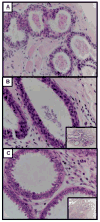
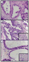
Results: Notation of FEA ranged from 0% to 35% of the cases interpreted, with most pathologists noting FEA on 4 or more test cases. At least one participant noted FEA in 34 of the 72 benign non-FEA cases. For the 6 reference FEA cases, participant agreement with the case reference FEA diagnosis ranged from 17% to 52%; diagnoses noted by participating pathologists for these FEA cases included columnar cell hyperplasia, usual ductal hyperplasia, atypical lobular hyperplasia, and atypical ductal hyperplasia.
Conclusions: We observed wide variation in the diagnosis of FEA among U.S. pathologists. This suggests that perceptions of diagnostic criteria and any implied risk associated with FEA may also vary. Surgical excision following a core biopsy diagnosis of FEA should be reconsidered and studied further.
Keywords: Atypia; Biopsy; Breast oncology; Flat epithelial atypia; Observer variability.
Copyright © 2017 Elsevier Ltd. All rights reserved.
Figures


Similar articles
-
Inter-observer variability between general pathologists and a specialist in breast pathology in the diagnosis of lobular neoplasia, columnar cell lesions, atypical ductal hyperplasia and ductal carcinoma in situ of the breast.Diagn Pathol. 2014 Jun 19;9:121. doi: 10.1186/1746-1596-9-121. Diagn Pathol. 2014. PMID: 24948027 Free PMC article.
-
Atypia on breast core needle biopsies: reproducibility and significance.Ann Clin Lab Sci. 2009 Summer;39(3):270-6. Ann Clin Lab Sci. 2009. PMID: 19667411
-
Pure flat epithelial atypia (DIN 1a) on core needle biopsy: study of 60 biopsies with follow-up surgical excision.Breast Cancer Res Treat. 2011 Jan;125(1):121-6. doi: 10.1007/s10549-010-1208-1. Epub 2010 Oct 14. Breast Cancer Res Treat. 2011. PMID: 20945087
-
Flat epithelial atypia of the breast: characteristics and behaviors.Am J Surg. 2011 Feb;201(2):245-50. doi: 10.1016/j.amjsurg.2010.02.009. Epub 2010 Sep 22. Am J Surg. 2011. PMID: 20864078 Review.
-
Atypia in breast pathology: what pathologists need to know.Pathology. 2022 Feb;54(1):20-31. doi: 10.1016/j.pathol.2021.09.008. Epub 2021 Dec 3. Pathology. 2022. PMID: 34872753 Review.
Cited by
-
Are Columnar Cell Lesions the Earliest Non-Obligate Precursor in the Low-Grade Breast Neoplasia Pathway?Curr Oncol. 2022 Aug 11;29(8):5664-5681. doi: 10.3390/curroncol29080447. Curr Oncol. 2022. PMID: 36005185 Free PMC article. Review.
-
Diagnostic interobserver variability of atypia assessment in columnar cell lesions among a group of expert breast pathologists in the United Kingdom and the Republic of Ireland, on behalf of the UK national coordinating committee for breast pathology.Histopathology. 2025 May;86(6):953-966. doi: 10.1111/his.15402. Epub 2024 Dec 20. Histopathology. 2025. PMID: 39704183 Free PMC article.
-
Second International Consensus Conference on lesions of uncertain malignant potential in the breast (B3 lesions).Breast Cancer Res Treat. 2019 Apr;174(2):279-296. doi: 10.1007/s10549-018-05071-1. Epub 2018 Nov 30. Breast Cancer Res Treat. 2019. PMID: 30506111 Free PMC article. Review.
-
Isolated Flat Epithelial Atypia: Upgrade Outcomes After Multidisciplinary Review-Based Management Using Excision or Imaging Surveillance.J Breast Imaging. 2023 Jul 22;5(5):575-584. doi: 10.1093/jbi/wbad049. eCollection 2023 Sep-Oct. J Breast Imaging. 2023. PMID: 37744722 Free PMC article.
-
Upgrade Rate of Pure Flat Epithelial Atypia Diagnosed at Core Needle Biopsy: A Systematic Review and Meta-Analysis.Radiol Imaging Cancer. 2021 Jan 22;3(1):e200116. doi: 10.1148/rycan.2021200116. eCollection 2021 Jan. Radiol Imaging Cancer. 2021. PMID: 33778758 Free PMC article.
References
-
- Ward CJ, VLG Risk Management and Medico-Legal Issues in Breast Cancer. Clin Obstet Gynecol. 2016;59(2):439–446. - PubMed
-
- Prowler VL, Joh JE, Acs G, et al. Surgical excision of pure flat epithelial atypia identified on core needle breast biopsy. Breast. 2014;23(4):352–356. - PubMed
-
- Silverstein M. Where's the outrage? J Am Coll Surg. 2009;208(1):78–79. - PubMed
-
- Silverstein M, Recht A, Lagios MD, et al. Special report: Consensus conference III. Image-detected breast cancer: state-of-the-art diagnosis and treatment. J Am Coll Surg. 2009;209(4):504–520. - PubMed
-
- Tavassoli FA, Devilee P. Pathology and genetics of tumours of the breast and female genital organs. IARC; 2003.
MeSH terms
Grants and funding
LinkOut - more resources
Full Text Sources
Other Literature Sources
Medical

