

Association of Condylar Bone Quality with TMJ Osteoarthritis
- PMID: 28476093
- PMCID: PMC5502961
- DOI: 10.1177/0022034517707515
Association of Condylar Bone Quality with TMJ Osteoarthritis
Abstract
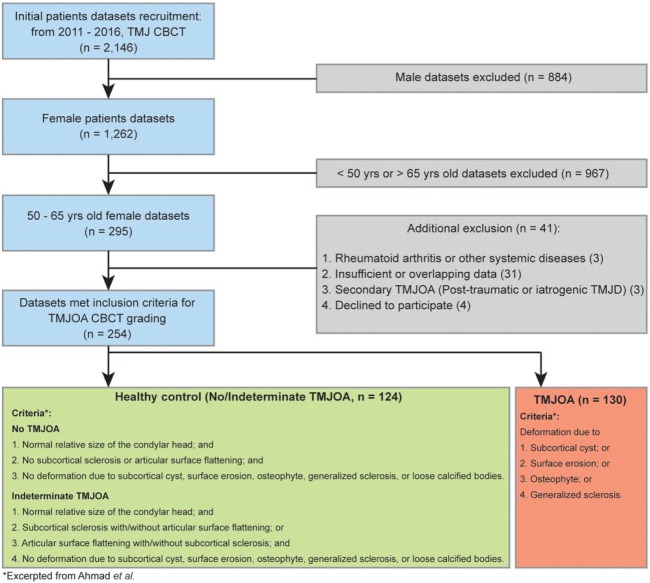
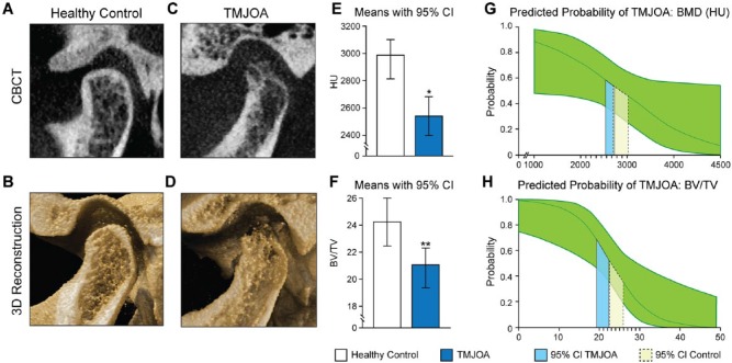
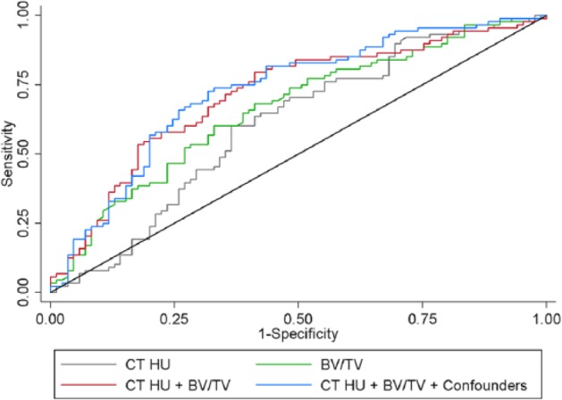
The etiology and treatment of temporomandibular joint (TMJ) osteoarthritis (TMJOA) remain complex and unclear. Based on clinical observations, we hypothesized that low condylar bone quality is significantly correlated with TMJOA and explored this association in a cross-sectional study with human patients. A total of 254 postmenopausal female participants were included in this study. Radiographic findings from cone beam computed tomography (CBCT) and clinical symptoms were used to classify each TMJ data sample as healthy control ( n = 124) or TMJOA ( n = 130). Condylar bone mineral density (BMD) (computed tomography Hounsfield unit [CT HU]) and bone volume fraction (BV/TV) were measured and modeled as predictors of healthy control versus TMJOA status in multilevel logistic regression analyses. Both CT HU (adjusted odds ratio [AOR] = 0.9989, interquartile odds ratio [IOR] = 0.4206) and BV/TV (AOR= 0.8096, IOR = 0.1769) were negatively associated with TMJOA ( P = 0.049, 0.011, respectively). To assess the diagnostic performance of CT HU and BV/TV for identification of TMJOA, receiver operating characteristic (ROC) curves were plotted. The estimated areas under the curve (AUC) were 0.6622 for BV/TV alone, 0.6074 for CT HU alone, and 0.7136 for CT HU and BV/TV together. The model incorporating CT HU and BV/TV together had a significantly higher AUC than the models using BV/TV alone ( P = 0.038) or HU alone ( P = 0.021). In conclusion, we found that low condylar bone quality was significantly correlated with TMJOA development and that condylar CT HU and BV/TV can be used together as a potential diagnostic tool for TMJOA. Careful clinical evaluation of the condyle coupled with appropriate radiographic interpretation would thus be critical for the early detection of TMJOA.
Keywords: bone volume; cone beam computed tomography; cross-sectional study; mandibular condyle; micro-computed tomography; multivariate analysis.
Conflict of interest statement
The authors declare no potential conflicts of interest with respect to the authorship and/or publication of this article.
Figures
References
-
- Agresti A. 2013. Categorical data analysis. Hoboken (NJ): John Wiley.
-
- Ahmad M, Hollender L, Anderson Q, Kartha K, Ohrbach R, Truelove EL, John MT, Schiffman EL. 2009. Research diagnostic criteria for temporomandibular disorders (RDC/TMD): development of image analysis criteria and examiner reliability for image analysis. Oral Surg Oral Med Oral Pathol Oral Radiol Endod. 107(6):844–860. - PMC - PubMed
-
- Akamatsu Y, Mitsugi N, Taki N, Takeuchi R, Saito T. 2009. Relationship between low bone mineral density and varus deformity in postmenopausal women with knee osteoarthritis. J Rheumatol. 36(3):592–597. - PubMed
-
- Akerman S, Kopp S, Rohlin M. 1988. Macroscopic and microscopic appearance of radiologic findings in temporomandibular joints from elderly individuals: an autopsy study. Int J Oral Maxillofac Surg. 17(1):58–63. - PubMed
MeSH terms
Grants and funding
LinkOut - more resources
Full Text Sources
Other Literature Sources
Medical
