. 2017 May 5;17(1):316.
doi: 10.1186/s12885-017-3301-x.

Gamma-glutamyltransferase activity in exosomes as a potential marker for prostate cancer

Affiliations

Gamma-glutamyltransferase activity in exosomes as a potential marker for prostate cancer

Kyojiro Kawakami et al. BMC Cancer. .

Abstract

Background: Exosomes or extracellular vesicles have the potential as a diagnostic marker for various diseases including cancer. In order to identify novel exosomal markers for prostate cancer (PC), we performed proteomic analysis of exosomes isolated from PC cell lines and examined the usefulness of the marker in patients.

Methods: Exosomes isolated by differential centrifugation from the culture medium of androgen-dependent LNCaP prostate cancer cell line and its sublines of partially androgen-independent C4, androgen-independent C4-2 and bone metastatic C4-2B were subjected to iTRAQ-based proteomic analysis. Exosomes were also isolated by immunocapture and separated by size exclusion chromatography and density gradient centrifugation. Protein expression was determined by Western blot analysis. GGT activity was measured using a fluorescent probe, γ-glutamyl hydroxymethyl rhodamine green (gGlu-HMRG). Immunohistochemical analysis of tissues was performed using anti-GGT1 antibody.

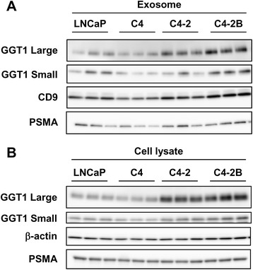
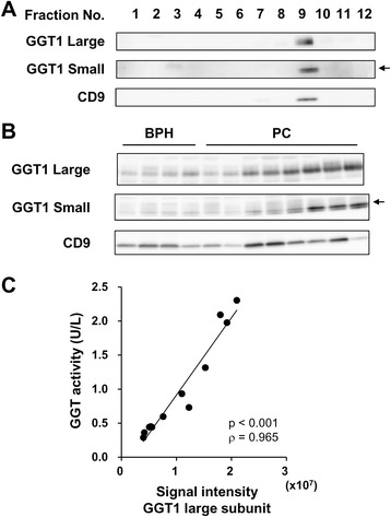
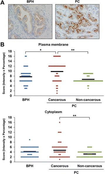
Results: Among proteins upregulated in C4-2 and C4-2B cells than in LNCaP cells, we focused on gamma-glutamyltransferase 1 (GGT1), a cell-surface enzyme that regulates the catabolism of extracellular glutathione. The levels of both GGT1 large and small subunits were elevated in exosomes isolated from C4-2 and C4-2B cells by differential centrifugation and by immunocapture with anti-CD9 or -prostate-specific membrane antigen (PSMA) antibody. In cell lysates and exosomes, GGT1 expression correlated with GGT activity. Size exclusion chromatography of human serum demonstrated the presence of GGT activity and GGT1 subunits in fractions positive for CD9. Density gradient centrifugation revealed the co-presence of GGT1 subunits with CD9 in exosomes isolated by differential centrifugation from human serum. Since GGT activity correlated with GGT1 expression in serum exosomes isolated by differential centrifugation, we measured serum exosomal GGT activity in patients. Unexpectedly, we found that serum exosomal GGT activity was significantly higher in PC patients than in benign prostatic hyperplasia (BPH) patients. In support of this finding, immunohistochemical analysis showed increased GGT1 expression in PC tissues compared with BPH tissues.

Conclusions: Our results suggest that serum exosomal GGT activity could be a useful biomarker for PC.

Keywords: Benign prostatic hyperplasia; Diagnostic marker; Exosome; Prostate cancer; γ-glutamyl transpeptidase; γ-glutamyltransferase 1.


Figures

Fig. 1
Fig. 1
GGT1 expression in exosomes isolated from PC cells by differential centrifugation. Exosomes were isolated from the culture medium of LNCaP, C4, C4–2 and C4–2B cells by differential centrifugation. Exosomes (a) and cell lysates (b) were subjected to Western blot analysis for GGT1 large and small subunits, CD9, PSMA and β-actin
Fig. 2
Fig. 2
GGT1 expression in exosomes isolated from PC cells by immunocapture. The culture medium of LNCaP, C4, C4–2 and C4–2B cells (30 mL) were incubated with magnetic beads (1 mg) conjugated with anti-CD9 (a) or -PSMA (b) antibody at 4 °C for 90 min. Whole immunocaptured exosomes were subjected to Western blot analysis for GGT1 large and small subunits, CD9, Alix and PSMA
Fig. 3
Fig. 3
GGT activity in cell lysates and exosomes isolated from PC cells by differential centrifugation. Cell lysates (a) and exosomes isolated from the culture medium of LNCaP, C4, C4–2 and C4–2B cells by differential centrifugation (b) were mixed with gGlu-HMRG. After incubation at room temperature for 1 h, fluorescence intensity (Ex/Em 490/520 nm) was measured by using microplate reader. GGT activity is shown as a percentage of LNCaP cells. *p < 0.05, compared with LNCaP cells, **p < 0.01, compared with C4 cells
Fig. 4
Fig. 4
GGT activity and GGT1 expression in exosomes isolated from human serum by SEC. Serum of a healthy individual (500 μL) was subjected to SEC. a CD9 expression in each fraction was measured by a sandwich ELISA. GGT activity in each fraction was determined by incubation with gGlu-HMRG at room temperature for 1 h and measurement of fluorescence intensity (Ex/Em 490/520 nm) using microplate reader. b The fractions 3–10 collected from a healthy individual were subjected to Western blot analysis for GGT1 large and small subunits and CD9. The upper band of the doublet corresponds to the GGT1 small subunit (shown by arrow)
Fig. 5
Fig. 5
GGT1 activity and GGT1 expression in exosomes isolated from human serum by differential centrifugation. a Exosomes isolated from serum of a healthy individual (500 μL) by differential centrifugation were separated by OptiPrep density gradient centrifugation. After ultracentrifugation, fractions were subjected to Western blot analysis for GGT1 large and small subunits and CD9. The upper band of the doublet corresponds to the GGT1 small subunit (shown by arrow). b Serum exosomes isolated by differential centrifugation from BPH (n = 4) and PC (n = 8) patients were subjected to Western blot analysis for GGT1 large and small subunits and CD9 as well as measurement of GGT activity using gGlu-HMRG. c Spearman’s rank correlation coefficient analysis was performed between the signal intensity of GGT1 large subunit and GGT activity
Fig. 6
Fig. 6
GGT activity in exosomes isolated by differential centrifugation from serum of PC and BPH patients. Exosomes were isolated by differential centrifugation from serum (210 μL) of PC (n = 31) and BPH (n = 8) patients. GGT activity was determined by incubation with gGlu-HMRG at room temperature for 1 h and measurement of fluorescence intensity (Ex/Em 490/520 nm) using microplate reader. Patient groups were compared for serum PSA concentration (a), serum GGT activity (b) and serum exosomal GGT activity (c). *p < 0.05, compared with BPH patients
Fig. 7
Fig. 7
Immunohistochemical analysis of GGT1 in PC and BPH tissues. Formalin-fixed paraffin-embedded biopsies and surgically resected tissue specimens from PC (n = 50) and BPH (n = 50) patients were stained for GGT1. a Representative images in BPH and PC tissues are shown. Original magnification ×400. b GGT1 expression on the plasma membrane and in the cytoplasm of cancerous and non-cancerous lesions of PC and BPH tissues is expressed as a score calculated by multiplying the intensity score with the percentage score. *p < 0.01, compared with BPH tissues. **p < 0.001, compared with the non-cancerous lesion

Similar articles

Cited by

References

    1. Colombo M, Raposo G, Thery C. Biogenesis, secretion, and intercellular interactions of exosomes and other extracellular vesicles. Annu Rev Cell Dev Biol. 2014;30:255–289. doi: 10.1146/annurev-cellbio-101512-122326. - DOI - PubMed
    1. Yoshioka Y, Kosaka N, Konishi Y, Ohta H, Okamoto H, Sonoda H, Nonaka R, Yamamoto H, Ishii H, Mori M, et al. Ultra-sensitive liquid biopsy of circulating extracellular vesicles using ExoScreen. Nat Commun. 2014;5:3591. doi: 10.1038/ncomms4591. - DOI - PMC - PubMed
    1. Melo SA, Luecke LB, Kahlert C, Fernandez AF, Gammon ST, Kaye J, LeBleu VS, Mittendorf EA, Weitz J, Rahbari N, et al. Glypican-1 identifies cancer exosomes and detects early pancreatic cancer. Nature. 2015;523(7559):177–182. doi: 10.1038/nature14581. - DOI - PMC - PubMed
    1. Siegel R, Ma J, Zou Z, Jemal A. Cancer statistics, 2014. CA Cancer J Clin. 2014;64(1):9–29. doi: 10.3322/caac.21208. - DOI - PubMed
    1. Kirby M, Hirst C, Crawford ED. Characterising the castration-resistant prostate cancer population: a systematic review. Int J Clin Pract. 2011;65(11):1180–1192. doi: 10.1111/j.1742-1241.2011.02799.x. - DOI - PubMed