. 2017 May 5;16(1):61.
doi: 10.1186/s12933-017-0544-4.

Dipeptidyl peptidase-4 (DPP-4) inhibition with linagliptin reduces western diet-induced myocardial TRAF3IP2 expression, inflammation and fibrosis in female mice

Affiliations

Dipeptidyl peptidase-4 (DPP-4) inhibition with linagliptin reduces western diet-induced myocardial TRAF3IP2 expression, inflammation and fibrosis in female mice

Annayya R Aroor et al. Cardiovasc Diabetol. .

Abstract

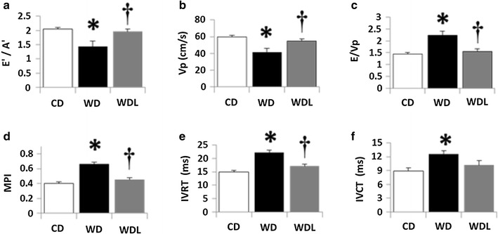
Background: Diastolic dysfunction (DD), a hallmark of obesity and primary defect in heart failure with preserved ejection fraction, is a predictor of future cardiovascular events. We previously reported that linagliptin, a dipeptidyl peptidase-4 inhibitor, improved DD in Zucker Obese rats, a genetic model of obesity and hypertension. Here we investigated the cardioprotective effects of linagliptin on development of DD in western diet (WD)-fed mice, a clinically relevant model of overnutrition and activation of the renin-angiotensin-aldosterone system.

Methods: Female C56Bl/6 J mice were fed an obesogenic WD high in fat and simple sugars, and supplemented or not with linagliptin for 16 weeks.

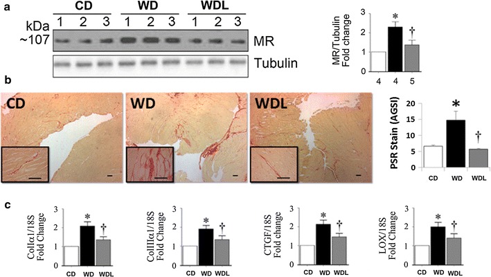
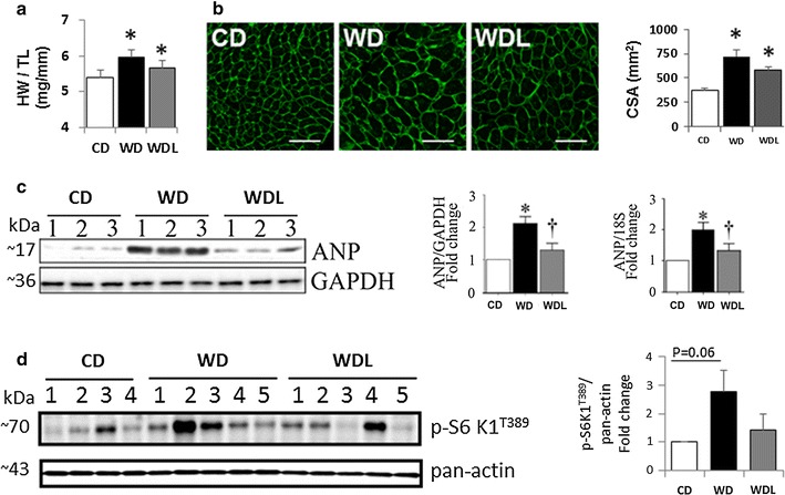
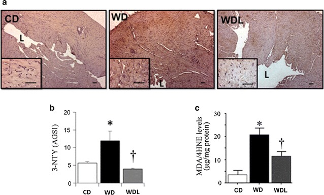
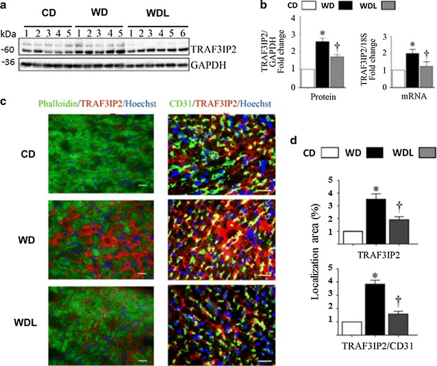
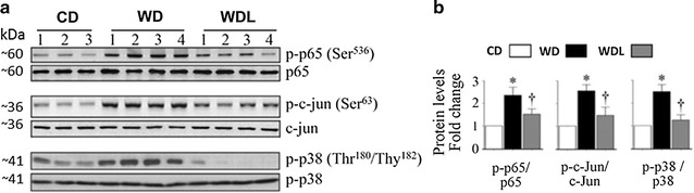
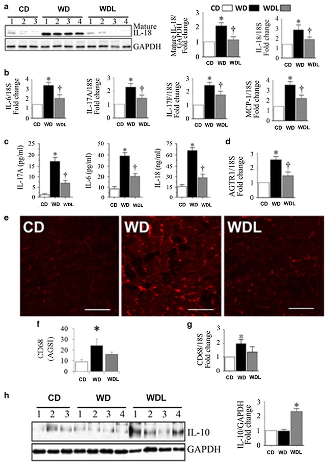
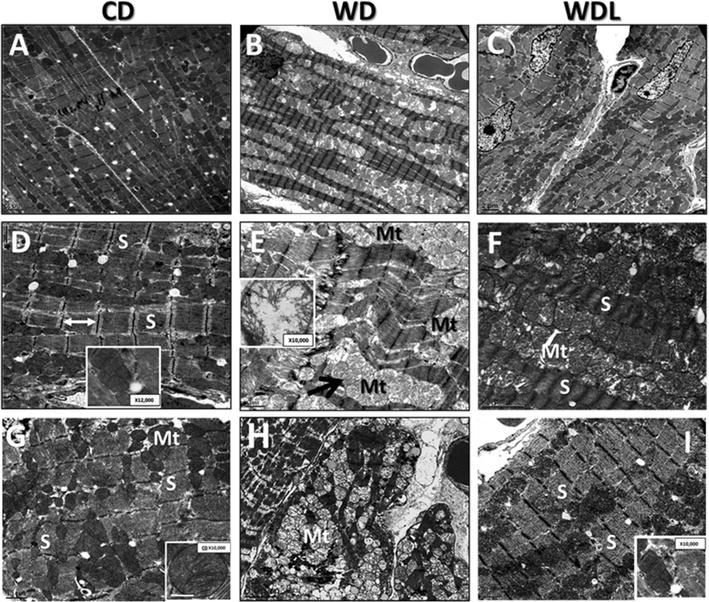
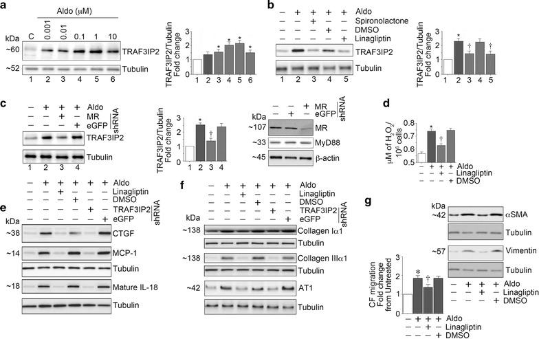
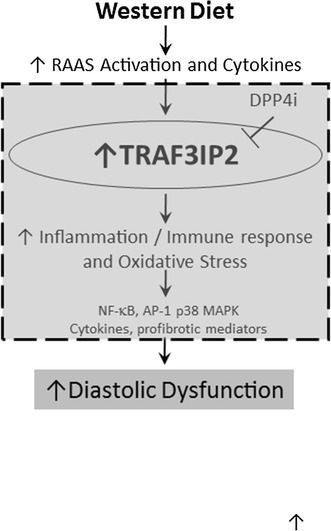
Results: WD induced oxidative stress, inflammation, upregulation of Angiotensin II type 1 receptor and mineralocorticoid receptor (MR) expression, interstitial fibrosis, ultrastructural abnormalities and DD. Linagliptin inhibited cardiac DPP-4 activity and prevented molecular impairments and associated functional and structural abnormalities. Further, WD upregulated the expression of TRAF3IP2, a cytoplasmic adapter molecule and a regulator of multiple inflammatory mediators. Linagliptin inhibited its expression, activation of its downstream signaling intermediates NF-κB, AP-1 and p38-MAPK, and induction of multiple inflammatory mediators and growth factors that are known to contribute to development and progression of hypertrophy, fibrosis and contractile dysfunction. Linagliptin also inhibited WD-induced collagens I and III expression. Supporting these in vivo observations, linagliptin inhibited aldosterone-mediated MR-dependent oxidative stress, upregulation of TRAF3IP2, proinflammatory cytokine, and growth factor expression, and collagen induction in cultured primary cardiac fibroblasts. More importantly, linagliptin inhibited aldosterone-induced fibroblast activation and migration.

Conclusions: Together, these in vivo and in vitro results suggest that inhibition of DPP-4 activity by linagliptin reverses WD-induced DD, possibly by targeting TRAF3IP2 expression and its downstream inflammatory signaling.

Keywords: Diastolic dysfunction; Linagliptin; Myocardial fibrosis; Obesity; TRAF3IP2.


Figures

Fig. 1
Fig. 1
Linagliptin prevents WD-induced diastolic dysfunction. Echocardiographic assessment of cardiac function indicates that WD induces impairments in a E’/A’ tissue Doppler index of diastolic function, b Vp, flow propagation velocity of early mitral inflow; c E/Vp, an index of LV filling pressure; d MPI myocardial performance index of global cardiac function; e IVRT isovolumic relaxation time; and f IVCT isovolumic contraction time. These functional impairments are prevented by administration of linagliptin. *p < 0.05 CD vs WD;p < 0.05 WD vs WDL. N = 10/group
Fig. 2
Fig. 2
Linagliptin prevents WD-induced cardiac fibrosis. a WD induces profibrotic mineralocorticoid receptor (MR) protein expression and this effect is blunted by linagliptin. b Representative micrographs (4× with 40× inset) show interstitial fibrosis by picrosirius red (PSR) staining in the heart and semi-quantitative assessment of PSR staining. Scale bars 50 μm. c Bar graphs show quantitative analysis of RT-qPCR- generated mRNA results for Col Iα, Col IIIα1, CTGF and LOX expressed as fold change from baseline in the CD group. *p < 0.05 vs CD and p < 0.05 vs WD. N = 5–8/group
Fig. 3
Fig. 3
Linagliptin inhibits WD-induced cardiac hypertrophy and induction of prohypertrophic mediators. a The ratio of heart weight to tibia length (HW/TL). N = 10/group. b Micrographs show wheat germ agglutinin (WGA) staining in the myocardium and accompanying bar graph shows semi-quantitative assessment of cardiomyocyte cross-sectional area (CSA). Scale bars 50 μm. c Western blot shows myocardial ANP expression and accompanying bar graphs show quantitative analysis of protein and mRNA expression as fold change from baseline in the CD group. N = 3/group. d Western blot shows myocardial phospho-S6K1 (Thr389) and pan-actin expression and accompanying bar graph shows fold change in expression from CD. *p < 0.5 vs CD and p < 0.05 vs WD. N = 4–5/group
Fig. 4
Fig. 4
Linagliptin abrogates WD-induced oxidative stress. a Representative micrographs show myocardial 3-nitrotyrosine immunostaining at low (4×) and high (40×) magnification (inset). L indicates lumen of the left ventricle. b Bar graph shows semi-quantitative analysis of 3-nitrotyrosine staining expressed as average gray scale intensities (AGSI). c Bar graph shows myocardial levels of malondialdehyde MDA and its degradation product, 4-hydroxynonenal (4HNE). *p < 0.05 vs CD and p < 0.05 vs WD. N = 3–6/group
Fig. 5
Fig. 5
Linagliptin prevents WD-induced increases in TRAF3IP2 expression. a Western blot shows myocardial TRAF3IP2 expression and accompanying bar graphs b show quantitative analysis of protein and mRNA expression as fold change from baseline in the CD group. c Immunofluorescent localization of TRAF3IP2 in the myocardium. The left panels show co-localization of phalloidin (cardiomyocytes, green) and TRAF3IP2 (red). The right panels show colocalization of CD31 (endothelial cells,) and TRAF3IP2 (yellow). d Bar graphs show quantitative analysis of TRAF3IP2 immunofluorescence in the myocardium (top bar graph) and in the coronary endothelium (bottom bar graph) expressed relative to CD. *p < 0.05 vs CD and p < 0.05 vs WD. N = 5–6/group
Fig. 6
Fig. 6
Linagliptin prevents WD-induced increases in p-65, c-jun and p38-MAPK activation. a Western blots show myocardial phospho- and total p-65, c-jun and p-38 MAPK. Accompanying bar graphs show results of densitometric analysis b of protein expression as fold change from baseline in the CD group. WD induces activation of these proinflammatory proteins which is largely prevented by linagliptin. *p < 0.05 vs CD and p < 0.05 vs WD. N = 5–6/group
Fig. 7
Fig. 7
Linagliptin inhibits the cardiac and systemic inflammatory responses to western diet. a WD increases myocardial IL-18 protein and mRNA expression, b pro-inflammatory cytokine gene expression (1L-6, IL-17A, IL-17F and MCP-1), c plasma pro-inflammatory cytokine levels (IL-6, IL-17A, and IL-18), and d myocardial angiotensin type 1 receptor (AGTR1) gene expression. These WD-induced inflammatory responses are prevented by linagliptin e, f WD increases CD68 positive immunofluorescence in the heart, indicating enhanced macrophage infiltration. g WD also increased CD68 mRNA expression. Linagliptin tended to reduce CD68 protein and mRNA levels. h Linagliptin induces anti-inflammatory IL-10 expression. *p < 0.05 vs CD and p < 0.05 vs WD. N = 5–6/group. AVGI average grey scale intensities
Fig. 8
Fig. 8
Linagliptin inhibits ultrastructural abnormalities in WD-fed hearts. Panels A, D and G depict the normal organized appearance of sarcomeres (S) alternating with a row of intermyofibrillar mitochondria (Mt). WD induces abnormal remodeling of mitochondria and sarcomeres as depicted in Panels B, E and H. Inset in panel E shows Mt swelling and loss of cristae structure. Linagliptin prevented these WD-induced ultrastructural abnormalities (Panels C, F and I). Panels AC ×800: scale bar 2 μm. Panels BI ×2000; scale bar 0.5 or 1 μm
Fig. 9
Fig. 9
Linagliptin inhibits aldosterone (Aldo)-induced cardiac fibroblast activation and migration. The MR agonist, Aldo upregulated TRAF3IP2 expression in a dose-dependent manner (a) and pretreatment with the MR antagonist spironolactone and silencing MR each attenuated Aldo-induced TRAF3IP2 expression (b and c). Further, linagliptin inhibited Aldo-induced oxidative stress as evidenced by reduced H2O2 generation (d), and the induction of CTGF, MCP-1, and IL-18 (e). Moreover, linagliptin inhibited upregulation in extracellular matrix proteins collagens Iα1 and IIIα1, and AT1R (f). These results were recapitulated by TRAF3IP2 knockdown (e and f). Importantly, linagliptin inhibited CF activation and migration (g), the hallmarks of cardiac fibrosis. These in vitro experiments were performed at least three times, and a representative immunoblot is shown
Fig. 10
Fig. 10
Schematic illustrates a possible causal role of TRAF3IP2 in western diet induced oxidative stress, inflammation, fibrosis and diastolic dysfunction, and the efficacy of linagliptin in reducing these cardiac impairments. Area within the dotted grey box summarizes novel data presented in this investigation

Similar articles

Cited by

References

    1. Jia G, DeMarco VG, Sowers JR. Insulin resistance and hyperinsulinemia in diabetic cardiomyopathy. Nat Rev Endocrinol. 2015;12:144–153. doi: 10.1038/nrendo.2015.216. - DOI - PMC - PubMed
    1. Schilling JD, Mann DL. Diabetic cardiomyopathy: bench to bedside. Heart Fail Clin. 2012;8(4):619–631. doi: 10.1016/j.hfc.2012.06.007. - DOI - PMC - PubMed
    1. Peterson LR, Waggoner AD, Schechtman KB, Meyer T, Gropler RJ, Barzilai B, Davila-Roman VG. Alterations in left ventricular structure and function in young healthy obese women: assessment by echocardiography and tissue doppler imaging. J Am Coll Cardiol. 2004;43(8):1399–1404. doi: 10.1016/j.jacc.2003.10.062. - DOI - PubMed
    1. Peterson LR, Saeed IM, McGill JB, Herrero P, Schechtman KB, Gunawardena R, Recklein CL, Coggan AR, Demoss AJ, Dence CS, et al. Sex and type 2 diabetes: obesity-independent effects on left ventricular substrate metabolism and relaxation in humans. Obesity (Silver Spring) 2012;20(4):802–810. doi: 10.1038/oby.2011.208. - DOI - PMC - PubMed
    1. De Simone G, Devereux RB, Chinali M, Roman MJ, Barac A, Panza JA, Lee ET, Howard BV. Sex differences in obesity-related changes in left ventricular morphology: the strong heart study. J Hypertens. 2011;29(7):1431–1438. doi: 10.1097/HJH.0b013e328347a093. - DOI - PMC - PubMed

Publication types

MeSH terms