Review
. 2017 Sep;92(3):558-568.
doi: 10.1016/j.kint.2017.02.033. Epub 2017 May 3.

The third path of tubulointerstitial fibrosis: aberrant endothelial secretome

Affiliations
Review

The third path of tubulointerstitial fibrosis: aberrant endothelial secretome

Mark Lipphardt et al. Kidney Int. 2017 Sep.

Abstract

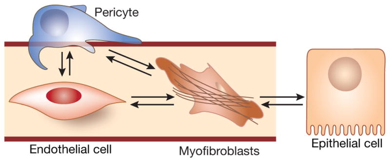
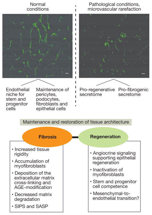
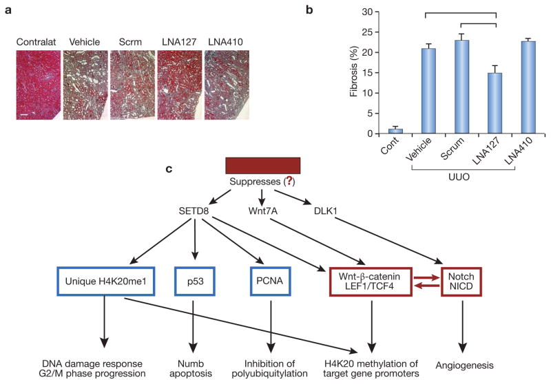
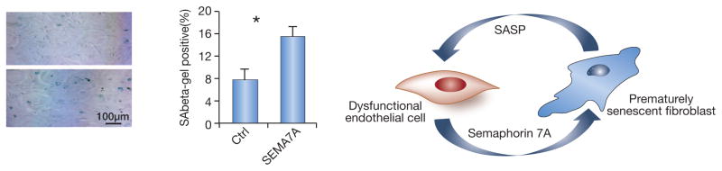
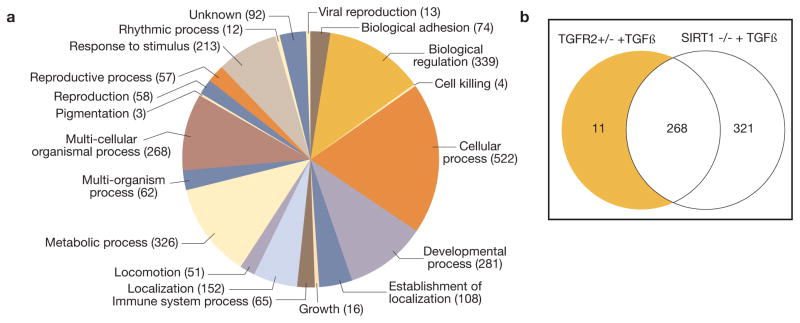
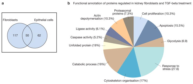
The secretome, defined as a portion of proteins secreted by specific cells to the extracellular space, secures a proper microenvironmental niche not only for the donor cells, but also for the neighboring cells, thus maintaining tissue homeostasis. Communication via secretory products exists between endothelial cells and fibroblasts, and this local mechanism maintains the viability and density of each compartment. Endothelial dysfunction, apart from obvious cell-autonomous defects, leads to the aberrant secretome, which predisposes fibroblasts to acquire a myofibroblastic fibrogenic phenotype. In our recent profiling of the secretome of such dysfunctional profibrogenic renal microvascular endothelial cells, we identified unique profibrogenic signatures, among which we detected ligands of Notch and Wnt-β-catenin pathways. Here, we stress the role of reprogramming cues in the immediate microenvironment of (myo)fibroblasts and the contribution of the endothelial secretome to the panoply of instructive signals in the vicinity of fibroblasts. We hope that this brief overview of endothelial-fibroblast communication in health and disease will lead to eventual unbiased proteomic mapping of individual secretomes of glomerular and tubular epithelial cells, pericytes, and podocytes through reductionist approaches to allow for the synthetic creation of a complex network of secretomic signals acting as reprogramming factors on individual cell types in the kidney. Knowledge of profibrogenic and antifibrogenic signatures in the secretome may garner future therapeutic efforts.

Keywords: endothelial cell; fibroblast; microenvironment; proteomics; reprogramming; secretome.


Figures

Figure 1
Figure 1. Four-compartmental model of (myo)fibroblast cross-talk with endothelial and epithelial cells. Grey background brackets the main subject of the present discussion
(Most recently, the fifth cell type, kidney-resident macrophages, closely associated with peritubular capillary bed, has been identified).
Figure 2
Figure 2
Endothelial secretome-dependent pathways for the maintenance and restoration of kidney architecture and pro-fibrogenic properties of aberrant endothelial secretome.
Figure 3
Figure 3. Profibrogenic effect of miR-127-3p
A and B: Masson’s trichrome staining of UUO kidneys in control mice, mice treated with the negative (scrambled) LNAs, and mice receiving complementary LNAs to miR-127 and miR-410. Ordinate shows the number of pixels corresponding to the detection of fibrotic areas. Horizontal lines above individual bars indicate p<0.05 between the corresponding groups. N=5 per each group of animals. Note that LNA to miR-127 mitigates fibrosis compared to both control groups, whereas LNA to miR-410 (another miR screen finding) is not effective, at least at the dose used. C: Targets of miR-127-3p. TargetScan identifies these target gene promoters relevant to Wnt/β-catenin and Notch pathways. Note a remarkable overlap with proteomic findings. In red are components of main focus.
Fig. 4
Fig. 4. Example of a dialog between the dysfunctional endothelium secreting semaphorin 7A and fibroblasts
A: Primary renal fibroblasts cultured in the presence of Sema-7A–Fc exhibit a 2-fold increase in prematurely senescent cells after only 48 h in culture. B: A cartoon depicting the vicious circle between dysfunctional endothelial secretome containing, among others, semaphorin 7A, and fibroblasts, which develop premature senescence and release SASP in turn affecting endothelial cells.
Figure 5
Figure 5
The secretome of cultured murine renal microvascular endothelial cells. A – categories of proteins detected in the secretome; B – a Venn diagram comparing the protein components of the secretome of endothelial cells isolated from SIRT1−/−, TGFR2+/−, and wild type mice and stimulated with TGF-β1.
Figure 6
Figure 6. Fibroblast and epithelial cells secretome
Human kidney fibroblast cell line and human epithelial cell line were grown to subconfluency. Medium was removed, and after washing in phosphate-buffered saline (PBS) the cells were incubated in serum-free DMEM. The cells were treated for 48 h with purified human TGFβ1 (5 ng/ml), and the supernatants were collected. The secretome proteins were prepared from the supernatant and separated by two-dimensional gel electrophoresis. The protein mapcompared to the control one and the differentially secreted proteins were identified by mass spectrometry.

References

    1. Mathivanan S, Ji H, Simpson RJ. Exosomes: extracellular organelles important in intercellular communication. J Proteomics. 2010;73:1907–20. - PubMed
    1. Mukherjee P, Mani S. Methodologies to decipher the cell secretome. Biochim Biophys Acta. 2013;1834:2226–32. - PMC - PubMed
    1. Rafii S, Butler JM, Ding BS. Angiocrine functions of organ-specific endothelial cells. Nature. 2016;529:316–25. - PMC - PubMed
    1. Ranganath SH, Levy O, Inamdar MS, et al. Harnessing the mesenchymal stem cell secretome for the treatment of cardiovascular disease. Cell Stem Cell. 2012;10:244–58. - PMC - PubMed
    1. Stamatiades EG, Tremblay ME, Bohm M, et al. Immune Monitoring of Trans-endothelial Transport by Kidney-Resident Macrophages. Cell. 2016;166:991–1003. - PMC - PubMed

MeSH terms