Review
. 2018 Jan;1860(1):22-39.
doi: 10.1016/j.bbamem.2017.04.028. Epub 2017 May 2.

Gating of Connexin Channels by transjunctional-voltage: Conformations and models of open and closed states

Affiliations
Review

Gating of Connexin Channels by transjunctional-voltage: Conformations and models of open and closed states

Thaddeus A Bargiello et al. Biochim Biophys Acta Biomembr. 2018 Jan.

Abstract

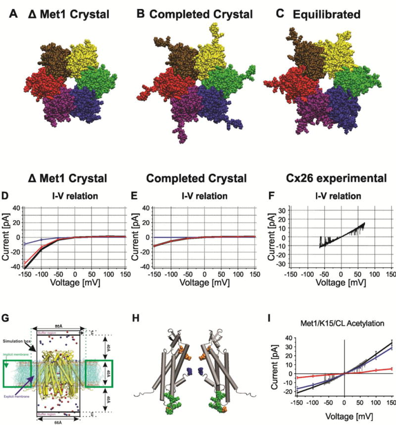
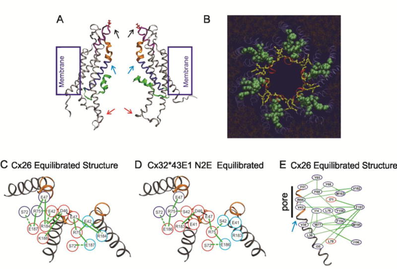
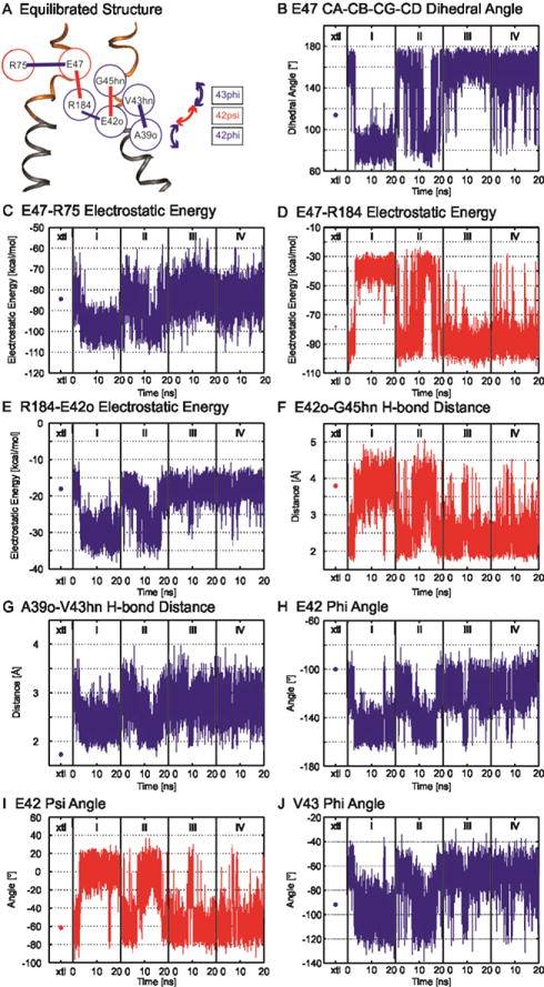
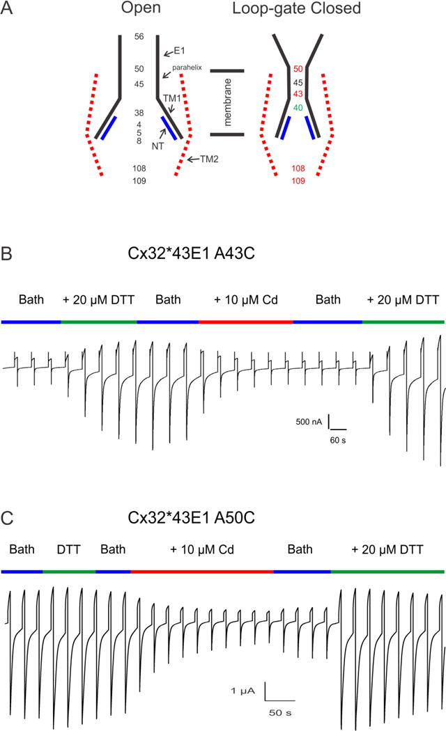
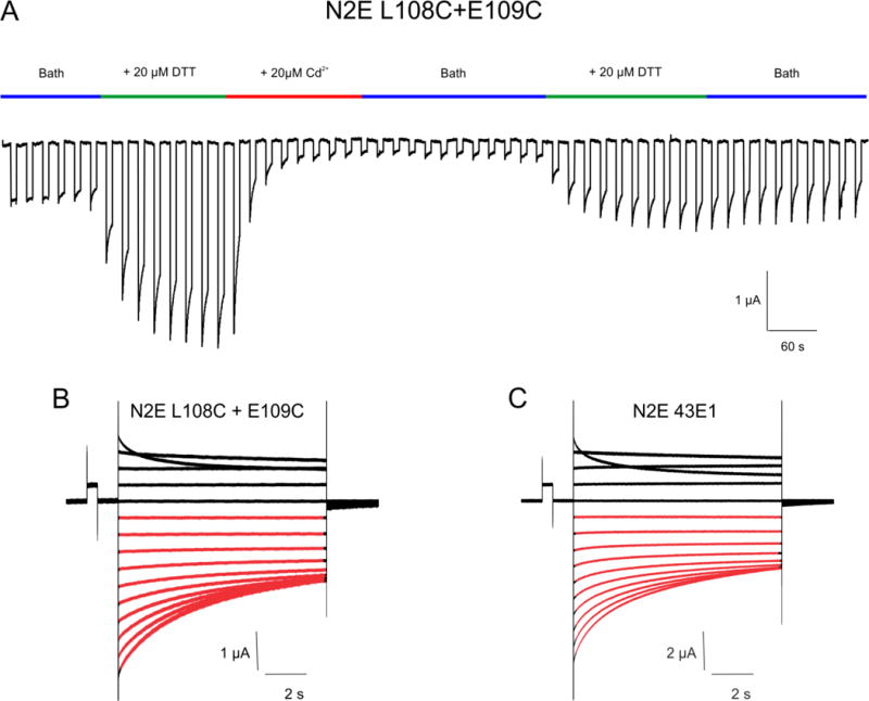
Voltage is an important physiologic regulator of channels formed by the connexin gene family. Connexins are unique among ion channels in that both plasma membrane inserted hemichannels (undocked hemichannels) and intercellular channels (aggregates of which form gap junctions) have important physiological roles. The hemichannel is the fundamental unit of gap junction voltage-gating. Each hemichannel displays two distinct voltage-gating mechanisms that are primarily sensitive to a voltage gradient formed along the length of the channel pore (the transjunctional voltage) rather than sensitivity to the absolute membrane potential (Vm or Vi-o). These transjunctional voltage dependent processes have been termed Vj- or fast-gating and loop- or slow-gating. Understanding the mechanism of voltage-gating, defined as the sequence of voltage-driven transitions that connect open and closed states, first and foremost requires atomic resolution models of the end states. Although ion channels formed by connexins were among the first to be characterized structurally by electron microscopy and x-ray diffraction in the early 1980's, subsequent progress has been slow. Much of the current understanding of the structure-function relations of connexin channels is based on two crystal structures of Cx26 gap junction channels. Refinement of crystal structure by all-atom molecular dynamics and incorporation of charge changing protein modifications has resulted in an atomic model of the open state that arguably corresponds to the physiologic open state. Obtaining validated atomic models of voltage-dependent closed states is more challenging, as there are currently no methods to solve protein structure while a stable voltage gradient is applied across the length of an oriented channel. It is widely believed that the best approach to solve the atomic structure of a voltage-gated closed ion channel is to apply different but complementary experimental and computational methods and to use the resulting information to derive a consensus atomic structure that is then subjected to rigorous validation. In this paper, we summarize our efforts to obtain and validate atomic models of the open and voltage-driven closed states of undocked connexin hemichannels. This article is part of a Special Issue entitled: Gap Junction Proteins edited by Jean Claude Herve.

Keywords: connexin; electrophysiology; homology models; metal bridging; molecular dynamics; voltage-gating.


Conflict of interest statement

The authors have no conflict of interests.

Figures

Figure 1
Figure 1
Single channel records illustrating two different voltage-dependent gating processes in Cx32*43E1 undocked hemichannels following expression in Xenopus oocytes. (A). Segment of a record of an outside-out patch containing a single channel illustrating Vj-gating transitions to three substates at a holding potential of −100 mV. (B). Cell attached recording of a single wild type channel at a holding potential of −70 mV illustrating loop-gating transitions. Note the slow time course of the gating transitions and that not all events lead to full channel closure. The records were obtained in presence of EDTA and EGTA, indicating that the two gating mechanisms do not depend on the presence of divalent cations. Reproduced from the Journal of General Physiology.
Figure 2
Figure 2
Structural models of Cx26 undocked hemichannels and corresponding computed currents-voltage relations with GCMC/BD. A–C. End on views of atomic models (A). The Cx26 crystal structure PDB ID:2ZW3. The larger pore diameter reflects the absence Met1 in the crystal structure. (B). The completed crystal structure in which all missing atoms were added to the crystal structure. The decreased pore diameter is a consequence of presence of Met1. (C). The structure of the average equilibrated atomic model following all-atom MD simulation in a fully hydrated POPC membrane. The large pore diameter is a consequence of the relaxation of the crystal structure. (D). I/V relation of the completed crystal structure with Met1 removed computed with GCMC/BD. Blue line is K+ current, red line is Cl current, black line is total current. (E). I/V relation of the completed crystal structure with Met1 included computed with GCMC/BD. Blue line is K+ current, red line is Cl current, black line is total current. (F) Single channel I/V relation of an excised (outside-out) undocked Cx26 hemichannel in symmetric 100mM KCl elicited by a ± 70 mV voltage ramp. (G). Schematic of the GCMC/BD simulation system. The connexin channel (yellow) embedded in explicit POPC lipid is inserted into an implicit membrane containing a circular hole. The explicit membrane prevents any leak current passing between the channel and implicit membrane. The upper compartment (extracellular part of the channel) was defined as the ground in voltage applications. 20 replicate 450-ns simulations were performed at each of seven voltages, ±150, ±100, ±50, and 0 mV, to plot the I/V relations. Blue circles, K+; red circles, Cl. (H) Positions of modified residues identified by Locke et al. (2009) that would alter the distribution of charge in the Cx26 channel pore, shown in a side view of two opposite subunits of the completed crystal structure. The positions of acetylated residues are colored as follows: blue, Met1; red, K15; green, K102, K103, K105, K108, K112, and K116 in CL/TM2; orange, γ-carboxyglutamated residues E42, E47, and E114. (I) The I/V relation of the MD equilibrated channel with Met/K15 6 cytoplasmic loop lysine residues acetylated. Blue line is K+ current, red line is Cl current, black line is total current. The experimental current trace depicted in gray is the current trace in panel F. Computed and experimental currents superimpose closely. Reproduced from the Journal of General Physiology.
Figure 3
Figure 3
Electrostatic and van der Waals networks stabilize the open state of Cx26 and N2E Cx32*43E1 undocked hemichannels. (A). Side view of the equilibrated Cx26 hemichannel. The parahelix (310 helix in Maeda et al.) which form the loop-gate permeability barrier in Cx32*43E1 and Cx50 hemichannels is depicted by the purple ribbon. The first transmembrane domain in blue. The TM1/E1 bend angle is marked by the turquoise arrow (it is in fact the TM1/parahelix bend angle). The first extracellular loop is colored blue. The N-terminus is colored green. The cytoplasmic entrance to the channel pore (109C) indicated by the red arrow. The extracellular entrance (residue 56) by the black arrow. (B) End on view from the extracellular entrance of the Cx26 hemichannel. Red ribbons are the parahelix, yellow rods are the electrostatic network, green spheres are the van der Waals network. (C). Schematic representation of the electrostatic network in the Cx26 equilibrated structure. (D). Schematic representation of the electrostatic network in the equilibrated N2E Cx32*43E1 hemichannel. (E). Schematic representation of the van der Waals network in the equilibrated atomic model of Cx26. Panels B–E are reproduced from Biophysical Journal.
Figure 4
Figure 4
Correlation map of the electrostatic network derived from correlated time series of electrostatic interactions present between two adjacent subunits. (A) Correlation map of electrostatic interactions. (Red and blue circles) Residues located in adjacent subunits. (B–J) Time series properties of specified interactions. (Blue) All interactions positively correlated with that of E47-R75; (red) those that are negatively correlated. Dots in column xtl are the energies of the interactions determined for the unequilibrated crystal structure. Additional details are provided in Kwon et al. [93]. Reproduced from Biophysical Journal.
Figure 5
Figure 5
(A). Schematic illustration of the Cx32*43E1 channel pore in the open and loop-gate closed state. Cysteine substitutions of the loci depicted in the open state were shown to be accessible to modification with thiol reagents. In the loop-gate closed state, cysteine substitutions at 43 and 50 form “high affinity” state-dependent Cd2+-thiolate metal bridges as does the double mutation 108C+109C. In contrast, the effect of Cd2+ on channels formed by individual mutations, 108C and 109C, is reversed by wash out of Cd2+. We attribute this effect to formation of low affinity bridges. Similarly, cysteine substitution at 40, are not accessible to MTS modification in the open state, and form “low affinity” metal bridges in the loop-gate closed state. Cysteine substitutions of G45C, interact with Cd2+ to lock the channel in the loop-gate closed state, but much of the effect can be reversed by wash-out of Cd2+ (incomplete reversal). Current relaxations of channels containing Q56C are unchanged by Cd2+. (B) High affinity Cd2+-thiolate bridge formation at 43C (see text). Activation of A43C currents requires application of either TPEN or DTT, as does restoration of currents following application of 1–10 μM Cd2+. (C) Currents from A50C channels differ from A43C in two ways, i) activation of currents does not require chelation, and ii) currents are partially restored by wash out of Cd2+, but full restoration requires application of metal chelators.
Figure 6
Figure 6
N2E Cx32*43E1 L108C +E109C undocked hemichannels are locked in loop-gate closed state by Cd2+. Currents were elicited by a train of voltage steps alternating between −10 and −70 mV, the time of reagent application is shown in the colored bars above the current trace. Low levels of initial currents are markedly increased by bath application of 20 μM DTT. Currents are decreased by application of 1–20 μM Cd2+ and are only partially restored by subsequent chelation of Cd2+ with 20 μM DTT. The result is consistent with formation of a high affinity Cd2+ site by substituted cysteines when the channel resides in the loop-gate closed state. (B and C). Currents from the N2E L108C+109C and “wild-type” N2E 43E1 (N2ECx32*43E1) hemichannels elicited by sequential voltage steps from +40 to −120 mV from a holding potential of 0mV. Closure of loop-gates at inside negative potentials corresponds to current relaxations shown in red. Closure of Vj-gates at inside positive potentials corresponds to current relaxations shown in black.

References

    1. Beyer EC, Berthoud VM. The Family of Connexin Genes. In: Harris AL, Locke D, editors. Connexins. Humana Press; 2009. pp. 3–26.
    1. Dewey MM, Barr L. Intercellular Connection between Smooth Muscle Cells: the Nexus. Science. 1962;137:670–672. - PubMed
    1. Revel JP, Yee AG, Hudspeth AJ. Gap Junctions between Electrotonically Coupled Cells in Tissue Culture and in Brown Fat. Proc Natl Acad Sci U S A. 1971;68:2924–2927. - PMC - PubMed
    1. Brightman MW, Reese TS. JUNCTIONS BETWEEN INTIMATELY APPOSED CELL MEMBRANES IN THE VERTEBRATE BRAIN. J Cell Biol. 1969;40:648–677. - PMC - PubMed
    1. Cohen-Salmon M, Ott T, Michel V, Hardelin JP, Perfettini I, Eybalin M, Wu T, Marcus DC, Wangemann P, Willecke K, Petit C. Targeted ablation of connexin26 in the inner ear epithelial gap junction network causes hearing impairment and cell death. Current biology: CB. 2002;12:1106–1111. - PMC - PubMed

Publication types

LinkOut - more resources