Review
. 2018 Feb:48:18-26.
doi: 10.1016/j.semcancer.2017.04.017. Epub 2017 May 2.

Protein kinase C as a tumor suppressor

Affiliations
Review

Protein kinase C as a tumor suppressor

Alexandra C Newton. Semin Cancer Biol. 2018 Feb.

Abstract

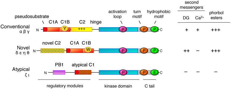
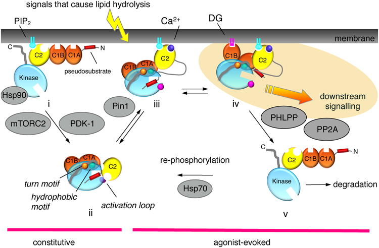
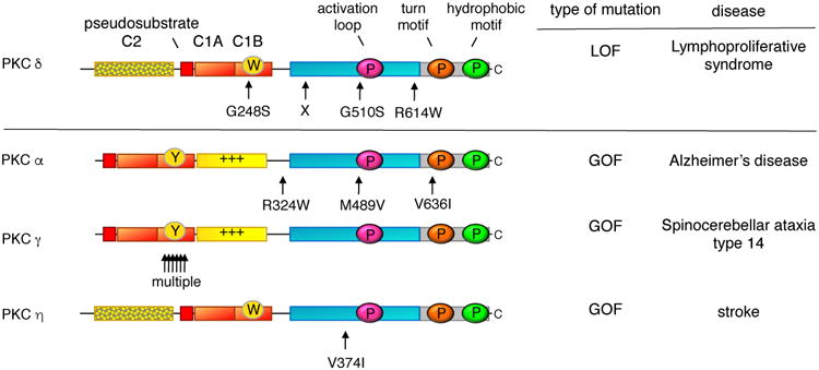
Protein kinase C (PKC) has historically been considered an oncoprotein. This stems in large part from the discovery in the early 1980s that PKC is directly activated by tumor-promoting phorbol esters. Yet three decades of clinical trials using PKC inhibitors in cancer therapies not only failed, but in some cases worsened patient outcome. Why has targeting PKC in cancer eluded successful therapies? Recent studies looking at the disease for insight provide an explanation: cancer-associated mutations in PKC are generally loss-of-function (LOF), supporting an unexpected function as tumor suppressors. And, contrasting with LOF mutations in cancer, germline mutations that enhance the activity of some PKC isozymes are associated with degenerative diseases such as Alzheimer's disease. This review provides a background on the diverse mechanisms that ensure PKC is only active when, where, and for the appropriate duration needed and summarizes recent findings converging on a paradigm reversal: PKC family members generally function by suppressing, rather than promoting, survival signaling.

Keywords: Diacylglycerol; LOF; PKC; Phorbol esters; Tumor suppressor.


Figures

Figure 1
Figure 1. Domain composition of PKC isozymes grouped by subfamily
All PKC isozymes comprise an N-terminal regulatory moiety that contains an autoinhibitory pseudosubstrate segment (red) that is immediately followed by a C1A domain (orange) and a C-terminal catalytic moiety. Conventional and novel PKC isozymes have a second C1 domain, the C1B domain (orange), which is the predominant diacylglycerol sensor in the full-length protein; its affinity for diacyglycerol is two orders of magnitude higher in the novel C1B domain compared to the conventional C1B domain because of a Trp (vs Tyr in conventional isozymes) at a site that toggles the affinity of the C1B domain for diacylglycerol (W vs Y indicated on domain). Conventional PKC isozymes have a Ca2+-binding C2 domain (yellow) that contains a basic surface (indicated by +++) that serves as a plasma membrane sensor via its recognition of PIP2. Novel PKC isozymes have a novel C2 domain that does not bind Ca2+ or lipids (mottled). Atypical PKC isozymes have a PB1 domain (purple) that mediates binding to protein scaffolds. The C-terminal kinase moiety contains the catalytic domain that has a priming phosphorylation site by PDK-1 (pink cirle) and a C-terminal tail (C tail; grey) that is phosphorylated at the turn motif (orange circle) and hydrophobic motif (green circle); atypical PKC isozymes have a Glu at the phosphoacceptor site of the hydrophobic motif. The sensitivity to second messengers, diacyglycerol (DG) and Ca2+, and to phorbol esters is shown on the right (+, ++, and +++ indicate relative affinity for C1 domain ligands).
Figure 2
Figure 2. Cartoon showing the multiple inputs that regulate the signaling lifetime of a conventional PKC
Following its biosynthesis, PKC is in an open, and degradation-sensitive, conformation in which all its regulatory modules are unmasked (species i). It is processed by a series of ordered phosphorylations that depend on the binding of Hsp90, with Cdc37, to a conserved PXXP motif in the kinase domain, the kinase complex mTORC2, and PDK-1. Phosphorylation at three priming sites, the activation loop, and two sites on the C-terminal tail, the turn motif and the hydrophobic motif, promote PKC to adopt an autoinhibited conformation in which the Ca2+-sensing C2 domain (yellow) clamps the autoinhibitory pseudosubstrate segment (red) in the substrate-binding cavity of the kinase domain (cyan), and the diacyglycerol-sensing C1 domains (orange) become masked (species ii). Hydrolysis of PIP2 results in Ca2+-dependent recruitment of PKC to the plasma membrane via engagement of the C2 domain (species iii), where PKC binds its membrane-embedded ligand, diacylglycerol, via primarily the C1B domain (species iv). This active PKC phosphorylates downstream substrates, such as Ras, to suppress oncogenic signaling. The membrane-bound conformation of PKC is sensitive to dephosphorylation, with the first event being dephosphorylation of the hydrophoboic motif catalyzed by PHLPP; subsequent dephosphorylation by PP2A produces a fully dephosphorylated PKC that is shunted for degradation by a proteosomal pathway (species v). However, binding of Hsp70 to the dephosphorylated turn motif allows PKC to become rephosphorylated to sustain the signaling lifetime of the enzyme. Phorbol esters (not shown) bind the C1B domain with two-orders of magnitude higher affinity than diacylglycerol (highlighted in yellow) and are not readily metabolized, trapping PKC in the open, phosphatase-sensitive conformation and resulting in chronic loss, or down-regulation, of PKC. Novel PKC isozymes are regulated by similar mechanisms except their C2 domain does not function as a Ca2+ or plasma membrane sensor, resulting in tht localization of novel PKC isozymes primarily to the more abundant and diacylglycerol-rich Golgi membranes. Atypical PKC isozymes are activated upon binding to specific protein scaffolds that tether the pseudosubstrate out of the substrate-binding cavity. Proteins indicated in grey are key regulators of the steady-state levels of PKC: Hsp70, Hsp90, mTORC2, and PDK-1 function to increase the steady-state levels of PKC by permitting/catalyzing processing phosphorylations; Pin1 and the phosphatases PHLPP and PP2A function to decrease the steady-state levels of PKC by permitting/catalyzing the dephosphorylation of PKC. Targeting any of these proteins will disrupt the balance of PKC signaling.
Figure 3
Figure 3. Germline mutations in PKC associated with disease
LOF mutations that are causative in a proliferative disease are a hallmark of a bonafide tumor suppressor: indicated are the positions of such germline mutations that have been identified in several families with lymphoproliferative syndrome; X indicates position of biallelic splice mutation that results in no expression of protein. In contrast to LOF mutations, germline mutations that enhance the activity of PKC are associated with degenerative diseases: indicated are rare variants in PKCα that segregated with affected family members with late onset Alzheimer's Disease, the multiple mutations in PKCγ that are causative in spinocerebellar ataxia type 14, and a variant in PKCη that is associated with increased risk to stroke.

References

    1. Takai Y, et al. Unsaturated Diacylglycerol as a Possible Messenger for the Activation of Calcium-activated, Phospholipid-dependent Protein Kinase System. Biochem Biophys Res Comm. 1979;91:1218–1224. - PubMed
    1. Hokin MR, Hokin LE. Enzyme Secretion and the Incorporation of P32 into Phospholipids of Pancreas Slices. J Biol Chem. 1953;203:967–977. - PubMed
    1. Carrasco S, Merida I. Diacylglycerol, when simplicity becomes complex. Trends Biochem Sci. 2007;32(1):27–36. - PubMed
    1. Manning G, et al. The protein kinase complement of the human genome. Science. 2002;298(5600):1912–34. - PubMed
    1. Levin DE, et al. A Candidate Protein Kinase C Gene, PKC1, Is Required for the S. cerevisiae Cell Cycle. Cell. 1990;62:213–224. - PubMed

Publication types