. 2017 Jun 30;292(26):10835-10844.
doi: 10.1074/jbc.M117.783464. Epub 2017 May 5.

Insights into the catalysis of a lysine-tryptophan bond in bacterial peptides by a SPASM domain radical S-adenosylmethionine (SAM) peptide cyclase

Affiliations

Insights into the catalysis of a lysine-tryptophan bond in bacterial peptides by a SPASM domain radical S-adenosylmethionine (SAM) peptide cyclase

Alhosna Benjdia et al. J Biol Chem. .

Abstract

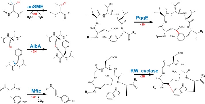
Radical S-adenosylmethionine (SAM) enzymes are emerging as a major superfamily of biological catalysts involved in the biosynthesis of the broad family of bioactive peptides called ribosomally synthesized and post-translationally modified peptides (RiPPs). These enzymes have been shown to catalyze unconventional reactions, such as methyl transfer to electrophilic carbon atoms, sulfur to Cα atom thioether bonds, or carbon-carbon bond formation. Recently, a novel radical SAM enzyme catalyzing the formation of a lysine-tryptophan bond has been identified in Streptococcus thermophilus, and a reaction mechanism has been proposed. By combining site-directed mutagenesis, biochemical assays, and spectroscopic analyses, we show here that this enzyme, belonging to the emerging family of SPASM domain radical SAM enzymes, likely contains three [4Fe-4S] clusters. Notably, our data support that the seven conserved cysteine residues, present within the SPASM domain, are critical for enzyme activity. In addition, we uncovered the minimum substrate requirements and demonstrate that KW cyclic peptides are more widespread than anticipated, notably in pathogenic bacteria. Finally, we show a strict specificity of the enzyme for lysine and tryptophan residues and the dependence of an eight-amino acid leader peptide for activity. Altogether, our study suggests novel mechanistic links among SPASM domain radical SAM enzymes and supports the involvement of non-cysteinyl ligands in the coordination of auxiliary clusters.

Keywords: biosynthesis; enzyme catalysis; enzyme mechanism; iron-sulfur protein; metalloenzyme; peptide biosynthesis; radical; radical AdoMet; radical SAM; radical SAM enzyme.


Conflict of interest statement

The authors declare that they have no conflicts of interest with the contents of this article

Figures

Figure 1.
Figure 1.
Reactions catalyzed by SPASM domain radical SAM enzymes. Shown are anSME (cysteine or serine oxidation into Cα-formylglycine), AlbA (thioether bond formation), MftC (oxidative decarboxylation), PqqE (C–C bond formation), and KW_cyclase (C–C bond formation).
Figure 2.
Figure 2.
Specificity of the KW_cyclase. a, gel electrophoresis analysis (SDS-PAGE, 12.5%) of the purified KW_cyclase. MW, molecular weight. b, UV-visible spectrum of KW_cyclase before (16 μm, dashed line) and after (18 μm, solid line) anaerobic reconstitution of the [Fe-S] clusters. c, sequences of the ME_30, ME_30AW, ME_30KF, and ME_30KA peptides. Black circles correspond to the putative peptide leader based on the alignment in Fig. 3a. The conserved residues are depicted as blue circles. Red circles indicate the residues that form the cyclic KGDGW peptide. The second KGDGW motif is depicted as purple circles. Mutated residues are indicated by a yellow circle. d, HPLC analysis of the reductive cleavage of SAM at time 0 (top trace) and after 180-min incubation with KW_cyclase (bottom trace) (see below for experimental conditions). Detection was performed at 257 nm, and 5′-dA was further analyzed by MS. e, HPLC analysis of the ME_30 peptide before (top trace) and after 180-min incubation with the KW_cyclase (bottom trace). Detection was performed at 280 nm (see below for experimental conditions). f, MALDI-TOF MS analysis of the various peptides used as substrate before (top red traces) and after (bottom blue traces) incubation with KW_cyclase. Reactions were performed by incubating the reconstituted KW_cyclase (50 μm) with peptide (1 mm), SAM (1 mm), DTT (3 mm), and sodium dithionite (2 mm) under anaerobic conditions. g, LC/MS-MS analysis of the ME_30 peptide before (top trace) and after (bottom trace) incubation with KW_cyclase.
Figure 3.
Figure 3.
Minimal substrate for KW_cyclase. a, sequence alignment of ME_30 peptide homologs found in various bacteria. The putative peptide leader sequence is highlighted in black. The conserved residues are highlighted in blue, whereas the residues involved in the cyclic KGDGW peptide are highlighted in red. The second KGDGW motif, found in S. thermophilus is highlighted in purple. St, S. thermophilus; Sm, S. mitis; Sa, S. agalactiae; Ll, L. lactis; Pa, P. aeruginosa. b, sequences of the ME_30, MK_21, and VK_14 peptides. c, MALDI-TOF MS analysis of MK_21 (left panel) and VK_14 (right panel) peptides before (top traces) or after (bottom traces) incubation with KW_cyclase (see below for experimental conditions). d, kinetic analysis of the reaction catalyzed by KW_cyclase in the presence of the ME_30 peptide. The data represent the mean ± S.D. of three independent reactions. See below for experimental conditions. e, kinetic analysis of the reaction catalyzed by KW_cyclase in the presence of the MK_21 peptide. Reactions were performed by incubating the reconstituted KW_cyclase (68 μm) with peptide substrate (1 mm), SAM (1 mm), DTT (3 mm), and sodium dithionite (2 mm) under anaerobic conditions. The data represent the mean ± S.D. of three independent reactions.
Figure 4.
Figure 4.
Investigation of the [4Fe-4S] clusters present in KW_cyclase. a, sequence alignment of the conserved cysteine residues present in KW_cyclase (KW_C), anSME, AlbA, PqqE, and MftC. The cysteine residues involved in the coordination of auxiliary cluster I (Aux I) and II (Aux II), according to the anSME structure, are highlighted in blue and red, respectively. Residues conserved among at least three sequences are highlighted in black. Amino acids occupying the position of Cys-261 (in anSME) are highlighted in gray. b, molecular phylogenetic analysis of representative SPASM domain radical SAM enzymes: KW_cyclase, anSME, AlbA, PqqE, and MftC. The evolutionary history was inferred by using the maximum likelihood method. The percentage of replicate trees in which the associated taxa clustered together in the bootstrap test is indicated next to the branches (1000 replicates). The SPASM domain radical SAM enzymes catalyzing protein modifications are highlighted in blue. SPASM domain radical SAM enzymes catalyzing thioether bond formation on peptides are highlighted in green. c, structural model of the KW_cyclase. SAM is depicted in green and colored by atom elements. Cysteine residues predicted to be involved in the coordination of the radical SAM [4Fe-4S] (Cys-117, Cys-121, and Cys-124) and the coordination of the auxiliary cluster I (Cys-347, Cys-365, and Cys-419) and auxiliary cluster II (Cys-406, Cys-409, Cys-415, and Cys-437) are indicated. d, gel electrophoresis analysis (SDS-PAGE, 12.5%) of the purified A3, C437A, C419A, and C406A mutants. MW, molecular weight. e, UV-visible spectrum of the wild-type (10 μm, top traces) and the A3 mutant (10 μm, bottom trace) before (blue traces) and after (red traces) 20-min incubation with sodium dithionite. OD, optical density. f, LC/MS analysis of the peptide MK_21 incubated with the WT or the A3, C437A, C419A, or C406A mutants. Reactions were performed by incubating the respective proteins (50 μm) after anaerobic reconstitution with the MK_21 peptide substrate (1 mm), SAM (1 mm), DTT (3 mm), and sodium dithionite (2 mm) for 2 h. g, LC/MS-MS analysis of the MK_21 peptide and the cyclic MK_21* peptide produced by the (WT) KW_cyclase. The characteristic ions are indicated. Similar fragmentation patterns were obtained for the C437A, C419A, and C406A mutants.

References

    1. Broderick J. B., Duffus B. R., Duschene K. S., and Shepard E. M. (2014) Radical S-adenosylmethionine enzymes. Chem. Rev. 114, 4229–4317 - PMC - PubMed
    1. Mehta A. P., Abdelwahed S. H., Mahanta N., Fedoseyenko D., Philmus B., Cooper L. E., Liu Y., Jhulki I., Ealick S. E., and Begley T. P. (2015) Radical S-adenosylmethionine (SAM) enzymes in cofactor biosynthesis: a treasure trove of complex organic radical rearrangement reactions. J. Biol. Chem. 290, 3980–3986 - PMC - PubMed
    1. Benjdia A., and Berteau O. (2016) Sulfatases and radical SAM enzymes: emerging themes in glycosaminoglycan metabolism and the human microbiota. Biochem. Soc. Trans. 44, 109–115 - PubMed
    1. Berteau O., and Benjdia A. (2017) DNA repair by the radical SAM enzyme spore photoproduct lyase: from biochemistry to structural investigations. Photochem. Photobiol. 93, 67–77 - PubMed
    1. Arnison P. G., Bibb M. J., Bierbaum G., Bowers A. A., Bugni T. S., Bulaj G., Camarero J. A., Campopiano D. J., Challis G. L., Clardy J., Cotter P. D., Craik D. J., Dawson M., Dittmann E., Donadio S., et al. (2013) Ribosomally synthesized and post-translationally modified peptide natural products: overview and recommendations for a universal nomenclature. Nat. Prod. Rep. 30, 108–160 - PMC - PubMed

Publication types

MeSH terms

Associated data