. 2017 Jun 30;292(26):10899-10911.
doi: 10.1074/jbc.M116.762229. Epub 2017 May 5.

Filaments and fingers: Novel structural aspects of the single septin from Chlamydomonas reinhardtii

Affiliations

Filaments and fingers: Novel structural aspects of the single septin from Chlamydomonas reinhardtii

Andressa P A Pinto et al. J Biol Chem. .

Abstract

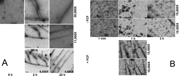
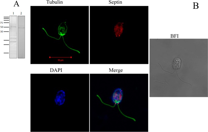
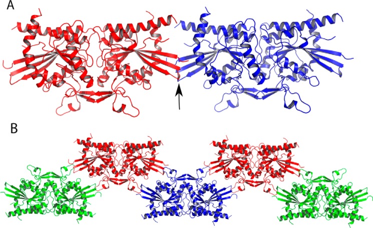
Septins are filament-forming GTP-binding proteins involved in many essential cellular events related to cytoskeletal dynamics and maintenance. Septins can self-assemble into heterocomplexes, which polymerize into highly organized, cell membrane-interacting filaments. The number of septin genes varies among organisms, and although their structure and function have been thoroughly studied in opisthokonts (including animals and fungi), no structural studies have been reported for other organisms. This makes the single septin from Chlamydomonas (CrSEPT) a particularly attractive model for investigating whether functional homopolymeric septin filaments also exist. CrSEPT was detected at the base of the flagella in Chlamydomonas, suggesting that CrSEPT is involved in the formation of a membrane-diffusion barrier. Using transmission electron microscopy, we observed that recombinant CrSEPT forms long filaments with dimensions comparable with those of the canonical structure described for opisthokonts. The GTP-binding domain of CrSEPT purified as a nucleotide-free monomer that hydrolyzes GTP and readily binds its analog guanosine 5'-3-O-(thio)triphosphate. We also found that upon nucleotide binding, CrSEPT formed dimers that were stabilized by an interface involving the ligand (G-interface). Across this interface, one monomer supplied a catalytic arginine to the opposing subunit, greatly accelerating the rate of GTP hydrolysis. This is the first report of an arginine finger observed in a septin and suggests that CrSEPT may act as its own GTP-activating protein. The finger is conserved in all algal septin sequences, suggesting a possible correlation between the ability to form homopolymeric filaments and the accelerated rate of hydrolysis that it provides.

Keywords: Chlamydomonas; GTPase; algae; arginine finger; crystal structure; filament; septin.


Conflict of interest statement

The authors declare that they have no conflicts of interest with the contents of this article

Figures

Figure 1.
Figure 1.
The structure of dimeric CrSEPT. A, schematic representation of the domain organization of CrSEPT, with the residue positions indicated. CrSEPT contains a polybasic region (P, yellow); the GTPase domain (orange), containing three of the five GTPase conserved motifs (G1 (red), G3 (green), and G4 (cyan) and a SUE (dark blue). B, schematic representation of the GTP-bound structure of CrSEPT. The conserved motifs are shown according to the color code established in A. C, structure-based sequence alignment of CrSEPT (PDB entry 5IRR) and other septin structures available (SEPT2 (2QNR), SEPT3 (3SOP), SEPT7 (3TW4), and SmSEPT10 (4KVA)). The elements of secondary structure are shown in the CrSEPT sequence in gray (β-strands) and green (α-helices). Important structural regions common to small GTPases are shown in transparent boxes, whereas septin-specific motifs (S1–S4) are shown within shaded boxes. In magenta are shown structural regions, which adopt an unusual conformation or are not normally observed when compared with other septins. Single residues that are highly conserved are marked in transparent boxes. Numbers indicate amino acid positions according to CrSEPT. The residue corresponding to the arginine finger is marked in red. *, basic residues that pack over the guanine base.
Figure 2.
Figure 2.
Biochemical characterization of CrSEPT. A, GTPγS binding affinity for CrSEPT was determined using ITC. Top, raw data (for GDP and GTPγS); bottom, best fit to the data for GTPγS. The binding isotherm has been fitted to a single-site binding model, yielding a Kd of 5.4 ± 0.3 μm (n = 0.84). GDP binding was not detectable (data not shown). B, influence of the nucleotide on the oligomeric state of CrSEPT. Aliquots of CrSEPT (10 μm), either nucleotide-free (solid line, monomer), complexed to GTPγS (dashed line, dimer), or complexed to GDP (dotted line) were analyzed by SEC on a Superdex 200 10/300 GL column and monitored at 280 nm. C, GTP hydrolysis by CrSEPT was assayed by measuring Pi release as a function of time. Initial rates were obtained from the slopes of phosphate-accumulation curves and fitted to a Michaelis-Menten model. Experiments were performed in triplicate, and the mean and S.D. values are reported. We obtained a kcat value of 2.41 ± 0.02 min−1. GraphPad Prism version 6.0 was used for all data fitting.
Figure 3.
Figure 3.
Comparison between the structures of SEPT2 and CrSEPT. A, schematic representation of SEPT2 (left, PDB entry 2QNR) and CrSEPT (right). In both structures, β1, β2, β3, α5′, α6, and the loop before the β hairpin (β7 and β8) are highlighted (in blue). The latter is indicated with an arrow. These are the most notable structural differences when compared with SEPT2, particularly the protrusion of the three β-strands. B, superimposition of CrSEPT, SEPT2 bound to Gpp(NH)p (PDB entry 3FTQ), and SEPT2 bound to GDP (PDB entry 2QNR). Side chains of CrSEPT Trp-163 and Trp-173 and SEPT2 Ile-88 and Leu-95 are shown. C, side-by-side close view of α6 region from CrSEPT (left) and GDP-bound SEPT2 (right). Black dashed lines highlight the interactions between the conserved His-195 and Arg-383 and neighbor residues, SEPT2 and CrSEPT, respectively. D, continuous electron density (gray mesh) is observed in the composite omit map for the switch I region. Residues at the beginning and end of the region are labeled explicitly, and the GTPγS is shown as solid spheres. E, schematic representation of the GTP-binding site generated with Coot using a method described by Clark and Labute (61). All residues within 4.5 Å of the ligand are represented, and the size of the halo around the residues represents the extent to which the solvent-accessible surface area was affected by ligand binding. Blue and green arrows indicate hydrogen bonds from main-chain and side-chain atoms, respectively. A π-anion interaction involving Arg-344 is explicitly represented.
Figure 4.
Figure 4.
GTPase Activity of CrSEPT employs a catalytic arginine finger. A, hydrogen-bond interaction between Arg-239 (chain B) and the γ-phosphate of GTPγS within the CrSEPT nucleotide-binding pocket of chain A. The Arg-239 also interacts via a hydrogen bond with Asp-185 (from the switch II region). The magnesium is shown as a sphere. B and C, influence of the nucleotide on the oligomeric state of the R239A and R239E mutants. The mutation to Ala (R239A) did not influence GTP-dependent dimerization, whereas substitution by Glu (R239E) completely prevented dimerization. D, both mutants were unable to hydrolyze GTP. E, GTPγS binding affinities for CrSEPT mutants were determined using ITC, as in Fig. 2A. The following Kd values were obtained from the fits: R239A, 7.6 ± 0.4 μm; R239E, 103 ± 4 μm; WT, 5.4 ± 0.3 μm. Experiments were performed in triplicate, and the mean and S. D. values are reported.
Figure 5.
Figure 5.
CrSEPT assembles into filaments in vitro. Shown are transmission electron micrographs of negatively stained septins at low salt concentration. A, samples produced by dialysis were taken after three different time intervals: 0, 2, and 22 h; B, sample produced by dilution. Top, samples were taken at three different times: within the first minute and after 1 and 3 h, respectively, in the presence of DMSO (control). Bottom, samples were taken only after the first hour, in the presence of 70 μm FCF. Nominal magnification for each panel is shown on the right.
Figure 6.
Figure 6.
CrSEPT immunodetection in Chlamydomonas. A, Western blot analysis was employed to examine the specificity of the anti-CrSEPT antibody to proteins extracted from Chlamydomonas cells. An extract of 10 ml of culture was prepared by using radioimmune precipitation buffer (50 mm Tris-HCl, pH 8, 150 mm NaCl, 1% Nonidet P-40, 0.5% sodium deoxycholate, 0.1% SDS) (61). Lane 1, total proteins extracted after resolution by 15% SDS-PAGE and stained with Coomassie Blue; lane 2, developed nitrocellulose membrane. B, septins are present mainly at the base of the flagella of C. reinhardtii. Shown is a confocal optical section of C. reinhardtii cells labeled with anti-CrSEPT. The immunolocalization of septin is shown as a bright red signal. This figure shows a punctate distribution of septins in the cells, stronger at the base of the flagella. A background fluorescent signal is present in the entire cell due to autofluorescence. Cells were also stained with anti-tubulin (green) and DAPI (blue). Panel 4 shows overlaid images from panels 1–3. Panel 5 shows the bright-field image (BFI) of Chlamydomonas cells.
Figure 7.
Figure 7.
NC canonical septin interfaces are not observed in CrSEPT crystals. A, superposing individual CrSEPT monomers across the NC interface of a homofilament composed of SEPT2. The arrow shows a minimal clash between the β-turns formed by strands β2 and β3 of each monomer. B, arrangement of CrSEPT dimers in crystal packing, showing how elements important for forming a canonical NC interface are unavailable because they are involved in packing contacts that stabilize the crystal.

Similar articles

Cited by

References

    1. Hartwell L. H. (1971) Genetic control of the cell division cycle in yeast. IV. Genes controlling bud emergence and cytokinesis. Exp. Cell Res. 69, 265–276 - PubMed
    1. Ong K., Wloka C., Okada S., Svitkina T., and Bi E. (2014) Architecture and dynamic remodelling of the septin cytoskeleton during the cell cycle. Nat. Commun. 5, 5698. - PMC - PubMed
    1. Longtine M. S., DeMarini D. J., Valencik M. L., Al-Awar O. S., Fares H., De Virgilio C., and Pringle J. R. (1996) The septins: roles in cytokinesis and other processes. Curr. Opin. Cell Biol. 8, 106–119 - PubMed
    1. Kinoshita M. (2006) Diversity of septin scaffolds. Curr. Opin. Cell Biol. 18, 54–60 - PubMed
    1. Pan F., Malmberg R. L., and Momany M. (2007) Analysis of septins across kingdoms reveals orthology and new motifs. BMC Evol. Biol. 7, 103. - PMC - PubMed

Publication types

MeSH terms

LinkOut - more resources