

Cytotoxic potentials of biologically fabricated platinum nanoparticles from Streptomyces sp. on MCF-7 breast cancer cells
- PMID: 28476980
- PMCID: PMC8676093
- DOI: 10.1049/iet-nbt.2016.0040
Cytotoxic potentials of biologically fabricated platinum nanoparticles from Streptomyces sp. on MCF-7 breast cancer cells
Abstract
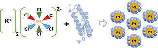
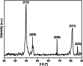
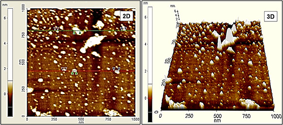
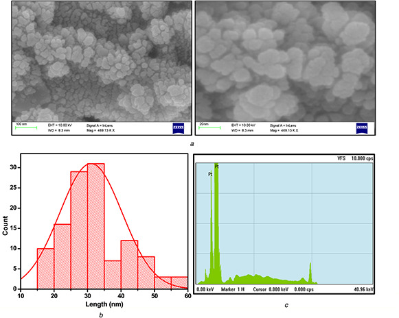
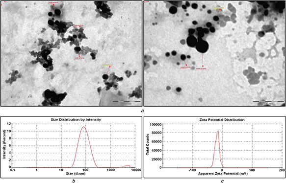
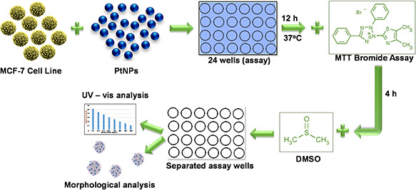
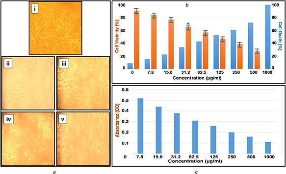
Biosynthesis of novel therapeutic nano-scale materials for biomedical and pharmaceutical applications has been enormously developed, since last decade. Herein, the authors report an ecological way of synthesising the platinum nanoparticles (PtNPs) using Streptomyces sp. for the first time. The produced PtNPs exhibited the face centred cubic system. The fourier transform infrared spectrum revealed the existence of amino acids in proteins which serves as an essential reductant for the formation of PtNPs. The spherical morphology of the PtNPs with an average size of 20-50 nm was observed from topographical images of atomic force microscopy and field emission scanning electron microscopy. The X-ray fluorescence spectrum confirms the presence of PtNPs with higher purity. The PtNPs size was further confirmed with transmission electron microscopy analysis and the particles were found to exist in the same size regime. Additionally, PtNPs showed the characteristic surface plasmon resonance peak at 262 nm. Dynamic light scattering studies report that 97.2% of particles were <100 nm, with an average particle diameter of about 45 nm. Furthermore, 3-(4, 5-dimethyl-2-thiazolyl)-2, 5-diphenyl-tetrazolium assay based in vitro cytotoxicity analysis was conducted for the PtNPs, which showed the inhibitory concentration (IC50) at 31.2 µg/ml against Michigan Cancer Foundation-7 breast cancer cells.
Figures

References
-
- Riggio C. Raffa V. Cuschieri A.: ‘Synthesis, characterisation and dispersion of zinc oxide nanorods for biomedical applications’, IET Micro Nano Lett., 2010, 5, (5), pp. 355 –360
-
- Reddy M.V. Tse L.Y. Bruce W.K.Z. et al.: ‘Low temperature molten salt preparation of nano‐SnO2 as anode for lithium‐ion batteries’, Mater. Lett., 2015, 138, pp. 231 –234
-
- Luximon A.B. Jhurry D.: ‘New avenues for improving pancreatic ductal adenocarcinoma (PDAC) treatment: selective stroma depletion combined with nano drug delivery’, Cancer Lett., 2015, 369, pp. 266 –273 - PubMed
-
- Li Y. Yang J. Song J.: ‘Nano‐energy system coupling model and failure characterization of lithium ion battery electrode in electric energy vehicles’, Renew. Sustain. Energy Rev., 2016, 54, pp. 1250 –1261
-
- Kumari A. Guliani A. Singla R. et al.: ‘Silver nanoparticles synthesised using plant extracts show strong antibacterial activity’, IET Nanobiotechnol., 2015, 9, (3), pp. 142 –152 - PubMed
MeSH terms
Substances
LinkOut - more resources
Full Text Sources
Other Literature Sources
