. 2017 Jun 27;8(26):42926-42938.
doi: 10.18632/oncotarget.17216.

Self-assembling nanoparticles encapsulating zoledronic acid inhibit mesenchymal stromal cells differentiation, migration and secretion of proangiogenic factors and their interactions with prostate cancer cells

Affiliations

Self-assembling nanoparticles encapsulating zoledronic acid inhibit mesenchymal stromal cells differentiation, migration and secretion of proangiogenic factors and their interactions with prostate cancer cells

Cinzia Borghese et al. Oncotarget. .

Abstract

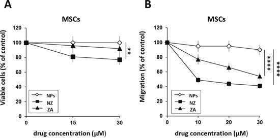
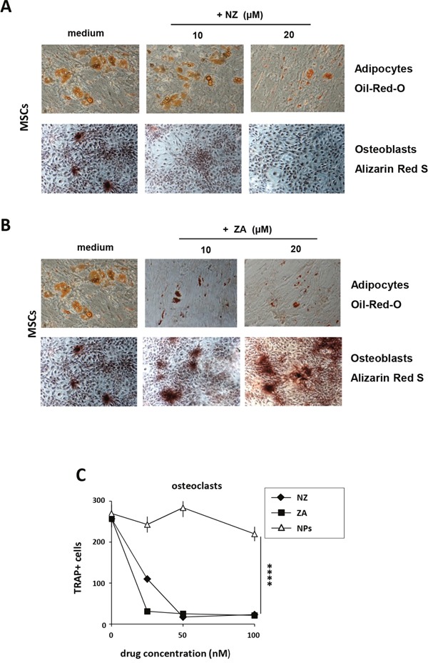
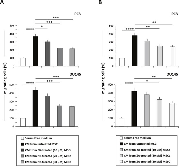
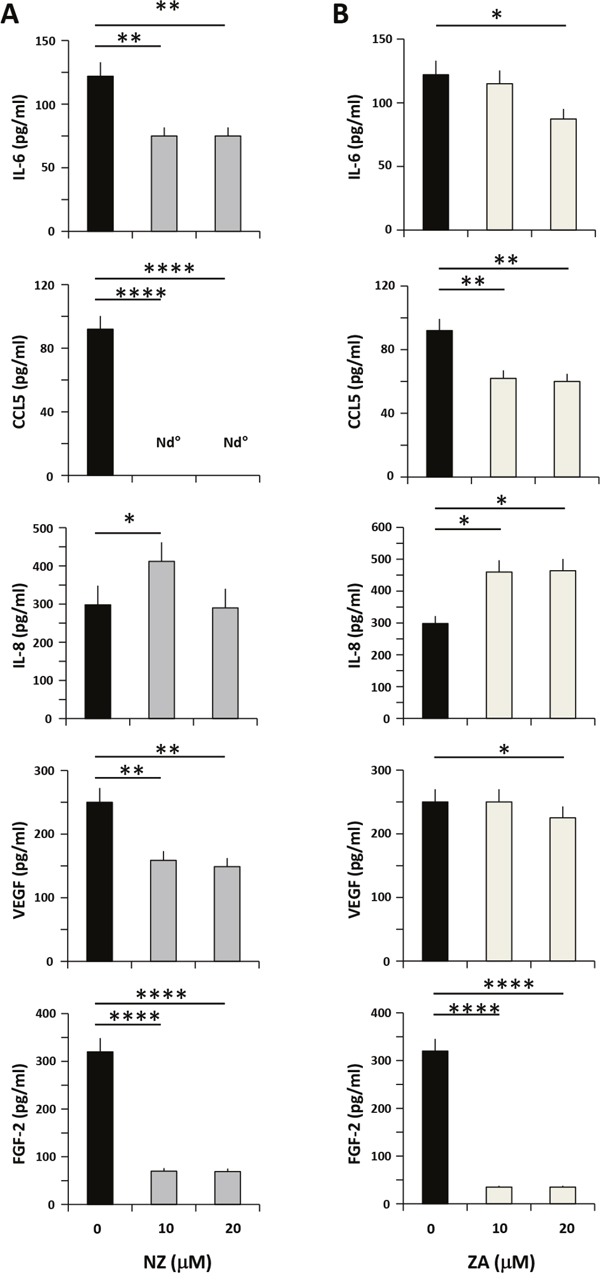
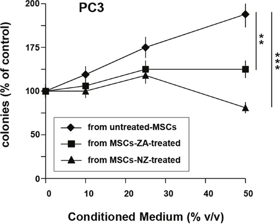
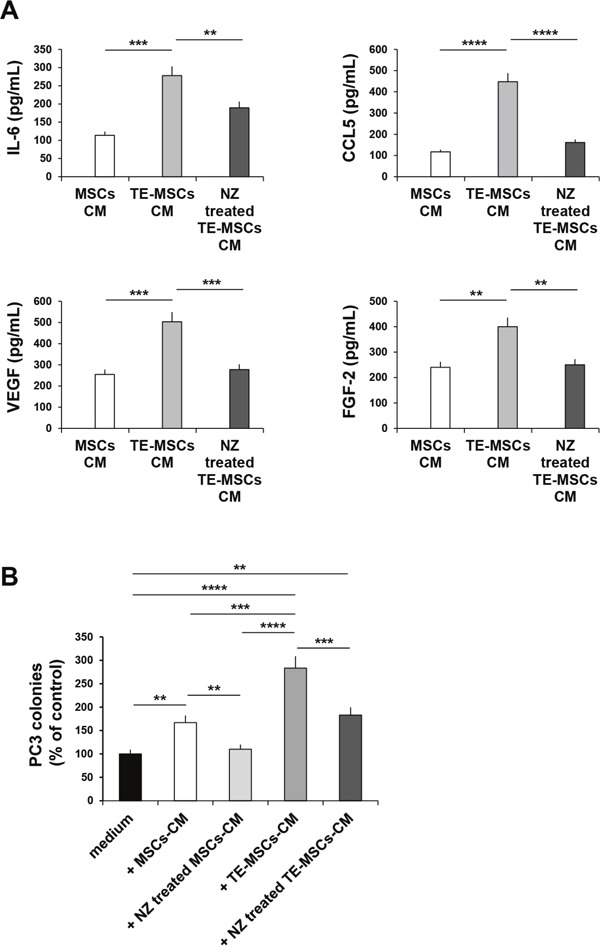
Zoledronic Acid (ZA) rapidly concentrates into the bone and reduces skeletal-related events and pain in bone metastatic prostate cancer (PCa), but exerts only a limited or absent impact as anti-cancer activity. Recently, we developed self-assembling nanoparticles (NPS) encapsulating zoledronic acid (NZ) that allowed a higher intratumor delivery of the drug compared with free zoledronic acid (ZA) in in vivo cancer models of PCa. Increasing evidence suggests that Bone Marrow (BM) Mesenchymal stromal cells (BM-MSCs) are recruited into the stroma of developing tumors where they contribute to progression by enhancing tumor growth and metastasis.We demonstrated that treatment with NZ decreased migration and differentiation into adipocytes and osteoblasts of MSCs and inhibited osteoclastogenesis. Treatment with NZ reduced the capability of MSCs to promote the migration and the clonogenic growth of the prostate cancer cell lines PC3 and DU145. The levels of Interleukin-6 and of the pro-angiogenic factors VEGF and FGF-2 were significantly reduced in MSC-CM derived from MSCs treated with NZ, and CCL5 secretion was almost totally abolished. Moreover, treatment of MSCs with supernatants from PC3 cells, leading to tumor-educated MSCs (TE-MSCs), increased the secretion of IL-6, CCL5, VEGF and FGF-2 by MSCs and increased their capability to increase PC3 cells clonogenic growth. Treatment with NZ decreased cytokine secretion and the pro-tumorigenic effects also of TE-MSCS. In conclusion, demonstrating that NZ is capable to inhibit the cross talk between MSCs and PCa, this study provides a novel insight to explain the powerful anticancer activity of NZ on PCa.

Keywords: mesenchymal stromal cells; prostate cancer; self-assembling nanoparticles; tumor microenvironment; zoledronic acid.


Conflict of interest statement

CONFLICTS OF INTEREST

The authors declare that they have no conflicts of interest.

Figures

Figure 1
Figure 1. Effect of NZ and ZA on MSCs proliferation and migration
(A) MSCs (2×10³) were cultured in the presence of increasing amounts of NZ, ZA or blank NPs (free liposomes). After 72 h, viable cells were evaluated by the MTT assay. (B) Migration through a collagen type I-coated Boyden chamber of MSCs (20 h) untreated and treated for 72 h with NZ, ZA or NPs (10, 20, 30 μM) in response to DMEM complete medium (10% FBS). Results are presented as percentage of migrated cells relative to control (untreated cells=100%). Values represent the mean ± SD of N=3 independent experiments.
Figure 2
Figure 2. Effect of NZ and ZA on osteoblast, adipocyte and osteoclast differentiation
Phase contrast microphotographs showing MSCs differentiation. MSCs were cultured with osteogenic (upper panel) or adipogenic (lower panel) medium alone or in the presence of (A) NZ or (B) ZA. Osteogenic differentiation was evaluated with Alizarin red staining (lower panel), adipogenic differentiation with Oil-Red-O staining (upper panel) (original magnification, 10×0.25). One representative experiment of three was reported. (C) Effect of NZ and ZA on osteoclastogenesis. Pre-OCs were incubated for 14 days with RANKL +M-CSF in the absence or in the presence of increasing concentrations of NZ, ZA or NPs. Then cells were stained for TRAP. Results represent the number of multinucleated TRAP-positive cells.
Figure 3
Figure 3. Treatment of MSCs with NZ or ZA decreased the migration of prostate cancer cells induced by MSCs-CM
Migration of PC3 and DU145 cells through a fibronectin-coated Boyden chamber in response to serum free medium (control=100%), CM from MSCs untreated or treated with (A) NZ or (B) ZA. Histograms represent the percentage of transmigrated cells after 20 h relative to control (cells migrated towards serum free medium). Values represent the mean ± SD of N=3 independent experiments.
Figure 4
Figure 4. Effects of NZ and ZA on cytokine/chemokine secretion by MSCs
MSCs were cultured in medium alone or with (A) NZ or (B) ZA for 72 h, then washed and incubated for additional 24 h in serum free medium. CM from MSCs was analyzed for IL-6, CCL5, IL-8, VEGF and FGF-2 secretion using specific ELISA assays. All samples were run in duplicate; supernatants from three different experiments were evaluated.
Figure 5
Figure 5. Treatment of MSCs with NZ or ZA decreased the clonogenic growth of PC3 cells induced by MSCs-CM
100 PC3 cells were plated in 24-well flat-bottomed plates and allowed to adhere for 24 h, then cultured in the presence of increased concentrations of supernatants from MSCs untreated or treated with ZA or NZ (20 μM). After 7 days, plates were observed under phase-contrast microscopy and colonies counted. Values represent the mean ± SD of N=3 independent experiments.
Figure 6
Figure 6. Effects of NZ on TE-MSCs
(A) BM-MSCs were cultured for 9 days with 10% of PC3-CM (MSCs become TE-MSCs). MSCs and TE-MSCs were cultured in medium alone or with NZ (10μM) for 72 h, then washed and incubated for additional 24 h in serum free medium. CM from MSCs and TE-MSCs treated or not with NZ were analyzed for IL-6, CCL5, VEGF and FGF-2 secretion using specific ELISA assays. All samples were run in duplicate; supernatants from three different experiments were evaluated. (B) 100 PC3 cells were plated in 24-well flat-bottomed plates and allowed to adhere for 24 h, then cultured in the presence of 20% CM from MSCs and TE-MSCs treated or not with NZ (10 μM). After 7 days plates were observed under phase-contrast microscopy and colonies counted. Values represent the mean ± SD of N=3 independent experiments.

Similar articles

Cited by

References

    1. Cammarota F, Laukkanen MO. Mesenchymal Stem/Stromal Cells in Stromal Evolution and Cancer Progression. Stem Cells Int. 2016;2016:4824573. - PMC - PubMed
    1. Poggi A, Musso A, Dapino I, Zocchi MR. Mechanisms of tumor escape from immune system: role of mesenchymal stromal cells. Immunol Lett. 2014;159:55–72. - PubMed
    1. Sun Z, Wang S, Zhao RC. The roles of mesenchymal stem cells in tumor inflammatory microenvironment. J Hematol Oncol. 2014;7:14–17. - PMC - PubMed
    1. Bergfeld SA, Blavier L, DeClerck YA. Bone marrow-derived mesenchymal stromal cells promote survival and drug resistance in tumor cells. Mol Cancer Ther. 2014;13:962–75. - PMC - PubMed
    1. Borghese C, Cattaruzza L, Pivetta E, Normanno N, De Luca A, Mazzucato M, Celegato M, Colombatti A, Aldinucci D. Gefitinib inhibits the cross-talk between mesenchymal stem cells and prostate cancer cells leading to tumor cell proliferation and inhibition of docetaxel activity. J Cell Biochem. 2013;114:1135–44. - PubMed

MeSH terms

LinkOut - more resources