Review
. 2018 Oct;41(4):985-998.
doi: 10.1007/s10143-017-0858-4. Epub 2017 May 6.

Multimodal use of indocyanine green endoscopy in neurosurgery: a single-center experience and review of the literature

Affiliations
Review

Multimodal use of indocyanine green endoscopy in neurosurgery: a single-center experience and review of the literature

Giuseppe Catapano et al. Neurosurg Rev. 2018 Oct.

Abstract

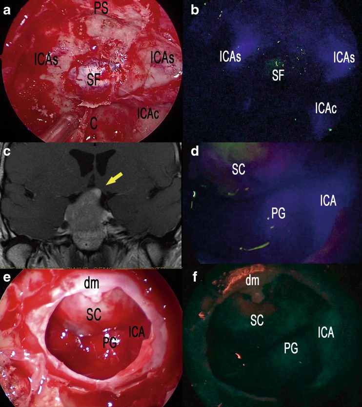
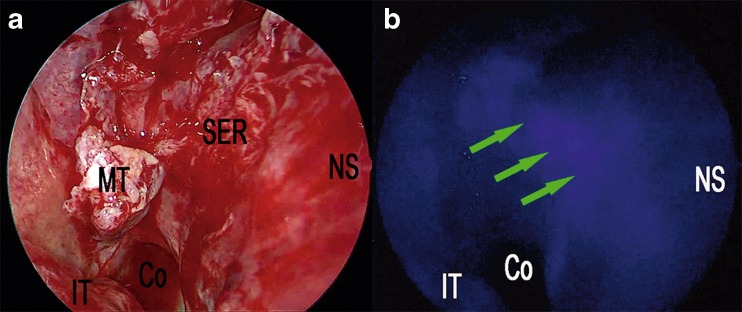
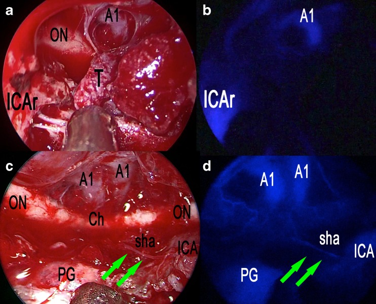
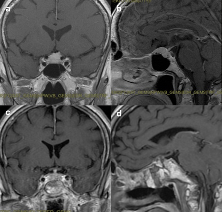
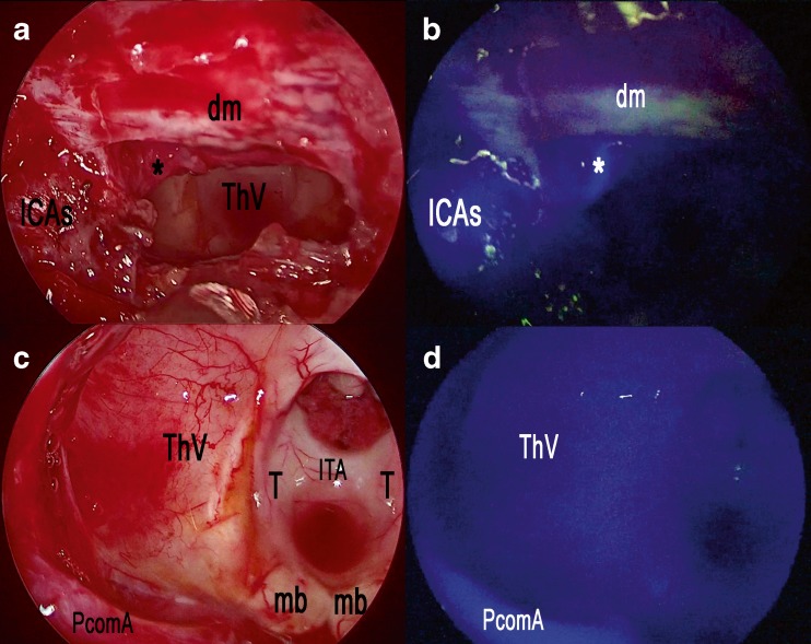
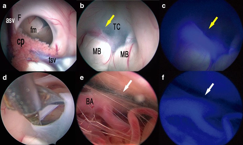
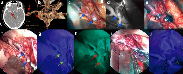
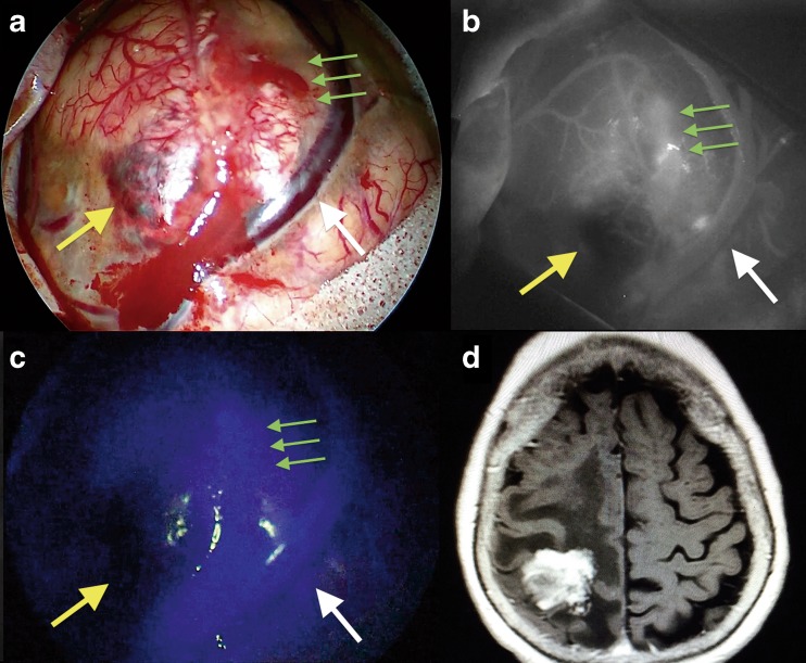
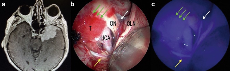
During the last 10 years, microscope-integrated indocyanine green fluorescence (m-ICG) has been widely used for assessing real-time blood flow during aneurysm surgery. More recently, an endoscope-integrated indocyanine green fluorescence (e-ICG) has been adopted as a versatile tool during different endoscopic neurosurgical procedures. The purpose of the present report is to evaluate multimodal applications of e-ICG during different endonasal, intraventricular, aneurysm and brain tumor surgeries and provide technical nuances. In addition, we reviewed the literature and identified and compare several overlapping case series of patients treated via an endoscopic integrated indocyanine green fluorescence technique. A total of 40 patients were retrospectively evaluated. Patients were divided into four main groups: (1) endoscopic endonasal approaches (n = 14); (2) ventricular endoscopic approach including patients undergoing third ventriculostomy (n = 8) and tumor biopsy (n = 1); (3) aneurysms surgery (n = 9); and (4) brain parenchymal tumors (n = 8). All patients were successfully treated using the e-ICG dynamic endoscopic visualization, and there were no perioperative complications. Such unique features open up a promising field of applications beyond the use of m-ICG in different surgical field due to the longer duration of e-ICG fluorescence up to 35 ± 7 min. E-ICG represents a new and effective technique for longer real-time visualization of vascular structures preserving normal tissues and functions during different transcranial and endonasal approaches. As the technology and e-ICG resolution improves, the technique has the potential to become a critical tool for different applications in neurosurgery.

Keywords: Aneurysm; Brain tumors; Endoscopy; Indocyanine green videoangiography; Third ventricle; Transsphenoidal.


Conflict of interest statement

Conflict of interest

The authors declare that they have no conflict of interest.

Ethical approval

All procedures performed in studies involving human participants were in accordance with the ethical standards of the institutional and/or national research committee and with the 1964 Helsinki Declaration and its later amendments or comparable ethical standards.

Informed consent

Informed consent was obtained from all individual participants included in the study.

Figures

Fig. 1
Fig. 1
Standard endoscopic endonasal approach to the sella. a The photo demonstrates the surgical field during sphenoidal step of the approach and the appearance under white light of the posterior wall of the sphenoid sinus. b The same surgical field as in a under near-infrared light after injection of ICG. c Post-contrast T1-weighted brain magnetic resonance imaging (MRI), coronal view, showing the pituitary macroadenoma with elevation and compression of the optic chiasm. The yellow arrow shows the position of the pituitary gland. d ICG fluorescence highlighting vascular structures within the intradural step of the approach. e The photo demonstrates the same field of view and the appearance of sellar area under white light. f The same surgical field under near-infrared light after injection of ICG using SPECTRA-A mode. SC suprasellar cistern, PG pituitary gland, SF sellar floor, C clivus, ICA internal carotid artery, ICAs parasellar segment of the internal carotid artery, ICAc clival segment of the internal carotid artery, dm dura mater, PS planum sphenoidale
Fig. 2
Fig. 2
Nasal step of extended endoscopic endonasal approach (a). Right nostril, harvesting of a pedicled nasal septal flap based on the posterior nasal septal artery (b). The same surgical field under near-infrared light after injection of ICG fluorescence highlighting vascular structures within nasal septum; the green arrows show the submucosal position of the posterior nasal septal artery. Co choana, IT inferior turbinate, MT middle turbinate, SER sphenoethmoid recess, NS nasal septum
Fig. 3
Fig. 3
Extended endoscopic endonasal transtuberculum/transplanum approach, removal of a tuberculum sellae meningioma. a The photo demonstrates the surgical field during tumor removal step and the appearance under white light. b The same surgical field as in a under near-infrared light after injection of ICG. c The photo demonstrates the surgical field after the complete excision of the tumor and the appearance under white light. d The same surgical field as in c under near-infrared light after injection of ICG. ON optic nerve, A1 anterior cerebral artery, T tumor, ICAr right internal carotid artery, Ch optic chiasm, PG pituitary gland, sha superior hypophyseal artery; green arrows show the course of the superior hypophyseal artery
Fig. 4
Fig. 4
Post-contrast T1-weighted brain MRI, coronal and sagittal view, showing preoperative (a, b) and postoperative (c, d) images of a tuberculum sellae meningioma
Fig. 5
Fig. 5
Extended endoscopic endonasal transtuberculum/transplanum approach, removal of a suprasellar craniopharyngioma. a The photo demonstrates the surgical field during the removal of the tumor and the appearance under white light. b The same surgical field as in c under near-infrared light after injection of ICG. c The photo demonstrates the surgical field after the complete removal of the tumor and the appearance under white light. d The same surgical field as in c under near-infrared light after injection of ICG. ICAs parasellar segment of the internal carotid artery, asterisk superior hypophyseal artery, dm dura mater, ThV third ventricle, T thalamus, ITA interthalamic adhesion, PcomA posterior communicating artery, mb mammillary body
Fig. 6
Fig. 6
Post-contrast T1-weighted brain MRI, coronal (a) and sagittal (b) views, showing a suprasellar craniopharyngioma with a large third ventricular extension
Fig. 7
Fig. 7
Right frontal endoscopic third ventriculostomy. a The photo demonstrates the surgical field in the lateral (a) and third (b) ventricles under white light. c The same surgical field as in b under near-infrared light after injection of ICG. d The photo demonstrates the surgical field during the opening of the third ventricle floor in the area of the tuber cinereum and the endoscopic exploration of the interpeduncular cistern (e) under white light. f The same surgical field as in e under near-infrared light after injection of ICG. asf anterior septal vein, cp choroid plexus, fm foramen of Monro, F fornix, tsv thalamostriate vein, MB mammillary body, BA basilar artery; yellow arrow shows the course of the posterior cerebral artery; white arrow shows the course of the basilar artery
Fig. 8
Fig. 8
Intraoperative findings during two middle cerebral artery aneurysms clipping cases via a right frontolateral approach. High-resolution computer tomography (CT) angiography and CT-based 3D reconstruction of two middle cerebral artery aneurysms (a, b); red arrows show the position of the aneurysms. c Right Sylvian fissure opening under microscopic view and white light; blue arrows show the position of the aneurysms. d The same surgical field as in c under indocyanine microscopic integrated view; blue arrows show the position of the aneurysms. e Clipping of the two middle cerebral aneurysms under microscopic view and white light. f Same case, right Sylvian fissure opening under endoscopic assisted view and white light; blue arrows show the position of the aneurysms. g The same surgical field as in f under near-infrared light after injection of ICG; green arrows show the position of the aneurysms. h The same surgical field as in g under near-infrared light after injection of ICG using SPECTRA-A mode; red arrows show the position of the aneurysms. i Clipping of the two middle cerebral aneurysms under endoscopic assisted view and white light. j Post-clipping step of the two middle cerebral aneurysms under endoscopic assisted view and near-infrared light after injection of ICG
Fig. 9
Fig. 9
The intraoperative findings during a parietal lobe metastasis of ovarian carcinoma removal via a right parietal approach. a Microscopic view before tumor removal under white light; yellow arrow shows the necrotic part of the lesion while the green arrows show the blood-brain barrier disruption. b The same surgical field as in a under indocyanine microscopic integrated view; yellow arrow shows the necrotic part of the lesion while the green arrows show the blood-brain barrier disruption. c The same surgical field as in b under endoscopic assisted view and near-infrared light after injection of ICG; yellow arrow shows the necrotic part of the lesion while the green arrows show the blood-brain barrier disruption
Fig. 10
Fig. 10
Intraoperative findings during spheno-orbital meningioma removal via a left pterional approach. Post-contrast T1-weighted brain MRI, axial view, showing the spheno-orbital meningioma extension. b Endoscopic assisted view showing the subfrontal exposure if the tumor under white light (a) and near-infrared light (c) after injection of ICG; yellow arrow shows a branch of the middle cerebral artery while the green arrows show the dural attachment of the lesion; the white arrow shows the a perforating artery over the left optic nerve

References

    1. Ben-Sira I, Riva CE. Fluorescein diffusion in the human optic disc. Investig Ophthalmol. 1975;14:205–211. - PubMed
    1. Betz CS, Zhorzel S, Schachenmayr H, Stepp H, Havel M, Siedek V, Leunig A, Matthias C, Hopper C, Harreus U. Endoscopic measurements of free-flap perfusion in the head and neck region using red-excited indocyanine green: preliminary results. J Plast Reconstr Aesthet Surg. 2009;62:1602–1608. doi: 10.1016/j.bjps.2008.07.042. - DOI - PubMed
    1. Bischoff PM, Flower RW. Ten years experience with choroidal angiography using indocyanine green dye: a new routine examination or an epilogue? Documenta ophthalmologica Advances in ophthalmology. 1985;60:235–291. doi: 10.1007/BF00157827. - DOI - PubMed
    1. Holm C, Tegeler J, Mayr M, Becker A, Pfeiffer UJ, Muhlbauer W. Monitoring free flaps using laser-induced fluorescence of indocyanine green: a preliminary experience. Microsurgery. 2002;22:278–287. doi: 10.1002/micr.10052. - DOI - PubMed
    1. Patel KM, Bhanot P, Franklin B, Albino F, Nahabedian MY. Use of intraoperative indocyanine-green angiography to minimize wound healing complications in abdominal wall reconstruction. J Plast Surg Hand Surg. 2013;47:476–480. - PubMed