. 2017 Oct;25(3-4):227-239.
doi: 10.1007/s10577-017-9556-x. Epub 2017 May 6.

Expression of progerin does not result in an increased mutation rate

Affiliations

Expression of progerin does not result in an increased mutation rate

Emmanuelle Deniaud et al. Chromosome Res. 2017 Oct.

Abstract

In the premature ageing disease Hutchinson-Gilford progeria syndrome (HGPS), the underlying genetic defect in the lamin A gene leads to accumulation at the nuclear lamina of progerin-a mutant form of lamin A that cannot be correctly processed. This has been reported to result in defects in the DNA damage response and in DNA repair, leading to the hypothesis that, as in normal ageing and in other progeroid syndromes caused by mutation of genes of the DNA repair and DNA damage response pathways, increased DNA damage may be responsible for the premature ageing phenotypes in HGPS patients. However, this hypothesis is based upon the study of markers of the DNA damage response, rather than measurement of DNA damage per se or the consequences of unrepaired DNA damage-mutation. Here, using a mutation reporter cell line, we directly compared the inherent and induced mutation rates in cells expressing wild-type lamin A or progerin. We find no evidence for an elevated mutation rate in progerin-expressing cells. We conclude that the cellular defect in HGPS cells does not lie in the repair of DNA damage per se.

Keywords: DNA damage; Lamin A; nuclear lamina; progeria.


Figures
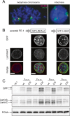
Fig. 1
Fig. 1
Expression of mutant and wild-type lamin As in MutaMouse cells. a FISH on metaphase chromosome spread (left) or interphase nucleus (right) from FE1 Muta Mouse cells hybridised with probes for phage λ DNA (red) and mouse chromosome 3 (green). DNA is counterstained with DAPI. b Immunofluorescence on parental FE1 cells (left column) and FE1 stable transfectants expressing wild-type (middle column) or Δ50 (right) mutant lamin A fused to GFP. Confocal mid planes showing GFP fluorescence (top row), immunofluorescence for laminA/C (middle row), merge (GFP/green, laminA/red and DAPI/blue) (bottom row). Scale bars = 5 μm. c Western blot of protein extracts from parental FE1 cells (left) and FE1 stable transfectants expressing wild-type (wt) or Δ50 mutant lamin A fused to GFP, and harvested at various passages. Membranes were probed with antibodies recognising GFP (top row), lamin A and lamin C (middle) and PCNA (bottom)—as a loading control
Fig. 2
Fig. 2
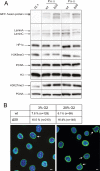
Histone modifications and nuclear morphology in lamin A-expressing cells. a Immunoblotting of proteins from FE-1 parental cells and from early (P20–25) and late (P50–55) passage FE-1 stable transfectants expressing wild-type (wt) or Δ50 mutant lamin A, with antibodies detecting laminA/C, HP1α, H3K9me3 and H3K27me3. H3 and PCNA serve as loading controls. b The table shows the proportion of abnormal nuclei scored in FE-1 transfectants expressing wild-type (wt) or Δ50 mutant GFP-tagged lamin A grown under conditions of low (3%) or high (20%) O2. n = number of nuclei scored. Below, confocal mid planes show examples of fields of Δ50-expressing cells grown under conditions of low (3%) or high (20%) O2. Green = GFP, blue = DAPI. Arrows indicate nuclei scored as abnormal. Scale bars = 5 μm
Fig. 3
Fig. 3
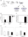
Determination of intrinsic mutant frequency in lamin A-expressing cells. a Schematic showing the determination of mutation frequency at λgt10lacZ sequences in FE-1 cells, by in vitro packaging of phage DNA and plating of infected E. coli on PGal selective plates. Only phage with mutations in lacZ (black filled) grow on selective PGal plates. Mutation frequency = ratio of pfu on PGal/pfu on non-selective (−PGal) plates. b and c Confirmation of efficacy of PGal selection. b The graph shows plating efficiency (pfu/ml in log scale) of wild-type (lacZ+) and known mutant (LacZ−) phage stocks on selective (+PGal, black bars) and non-selective (−PGal, white bars) plates. c Mutant frequency (pfu + PGal/pfu −PGal) measured for wild-type (white bars) and known mutant (LacZ−) stocks of phage. d Mutant frequency (×104) measured at the λgt10lacZ transgenes in FE-1 cells and in these cells stable expressing wild-type lamin A (wt) and HGPS mutant lamin A (Δ50). Cells were tested at low passage (<P24, left-hand graph) and then again at higher passage number (P = 48–53). The graphs show the mean ± s.e.m. for genomic DNAs isolated from two independent experiments, and with technical replicates for packaging of these DNAs
Fig. 4
Fig. 4
Induced mutant frequency in wild-type and mutant lamin A-expressing cells. a Mutant frequency (×104) measured at the λgt10lacZ transgenes in untreated control FE-1 cells and in cells exposed to 10 J/m2 UV 254 nm. b Mutant frequency (×104) in control and UV-treated FE-1 cells stably expressing wild-type lamin A (wt, white bars) or Δ50 mutant lamin A (black bars). Cells were tested at low passage (<P24, left-hand graph) and then again at a higher passage number (P = 48–53). The graphs show the mean ± s.e.m. for genomic DNAs isolated from two independent experiments, and with technical replicates for packaging of these DNAs. c Mutant frequency (×104) measured at the λgt10lacZ transgenes in control (DMSO) FE-1 cells and in cells exposed to 0.85 and 1.7 mM (100 and 200 μg/ml) ENU. d As in b but for control (DMSO) and 1.7 mM ENU-treated FE-1 cells stably expressing wt or Δ50 mutant lamin A
Fig. 5
Fig. 5
Indicators of the DNA damage response in wild-type and mutant lamin A-expressing cells. a Immunofluorescence with antibody detecting γH2A.X in FE-1 cells and cells exposed to 1 mM H2O2 for 25 min. In merge antibody is red, blue = DAPI. Scale bars = 5 μm. b Immunofluorescence with antibody detecting γH2A.X in FE-1 cells expressing wild-type (wt) or Δ50 mutant lamin A. In merge antibody is red, green = GFP, blue = DAPI. c Immunofluorescence with antibody detecting yH2A.X (red) in FE-1 cells expressing wild-type (wt) or Δ50 mutant lamin A. Green = GFP, blue = DAPI. Scale bars = 2 μm. d Immunoblot with antibodies detecting lamin A/C and γH2A.X in FE-1 cells and cells exposed to 1 mM H2O2 for 25 min. PCNA and H3 serve as loading controls. e Immunoblot with antibodies detecting lamin A/C and γH2A.X in FE-1 cells and transfectants expressing wild-type (wt) or Δ50 mutant lamin A

Similar articles

Cited by

References

    1. Agarwal AK, Fryns JP, Auchus RJ, Garg A. Zinc metalloproteinase, ZMPSTE24, is mutated in mandibuloacral dysplasia. Hum Mol Genet. 2003;12:1995–2001. doi: 10.1093/hmg/ddg213. - DOI - PubMed
    1. Barbie DA, Kudlow BA, Frock R, Zhao J, Johnson BR, Dyson N, Harlow E, Kennedy BK. Nuclear reorganization of mammalian DNA synthesis prior to cell cycle exit. Mol Cell Biol. 2004;24:595–607. doi: 10.1128/MCB.24.2.595-607.2004. - DOI - PMC - PubMed
    1. Bergo MO, Gavino B, Ross J, Schmidt WK, Hong C, Kendall LV, Mohr A, Meta M, Genant H, Jiang Y, et al. Zmpste24 deficiency in mice causes spontaneous bone fractures, muscle weakness, and a prelamin A processing defect. Proc Natl Acad Sci U S A. 2002;99:13049–13054. doi: 10.1073/pnas.192460799. - DOI - PMC - PubMed
    1. Boyle J, Kill IR, Parris CN. Heterogeneity of dimer excision in young and senescent human dermal fibroblasts. Aging Cell. 2005;4:247–255. doi: 10.1111/j.1474-9726.2005.00167.x. - DOI - PubMed
    1. Busuttil RA, Rubio M, Dolle ME, Campisi J, Vijg J. Oxygen accelerates the accumulation of mutations during the senescence and immortalization of murine cells in culture. Aging Cell. 2003;2:287–294. doi: 10.1046/j.1474-9728.2003.00066.x. - DOI - PubMed

Publication types

LinkOut - more resources