Review
. 2017 Oct;174(19):3211-3225.
doi: 10.1111/bph.13850. Epub 2017 Jun 7.

Cognitive deficits caused by prefrontal cortical and hippocampal neural disinhibition

Affiliations
Review

Cognitive deficits caused by prefrontal cortical and hippocampal neural disinhibition

Tobias Bast et al. Br J Pharmacol. 2017 Oct.

Abstract

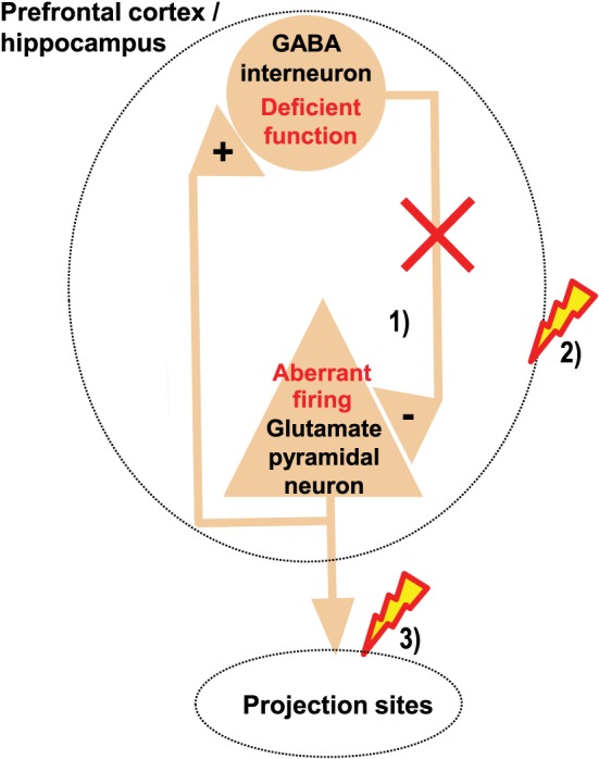
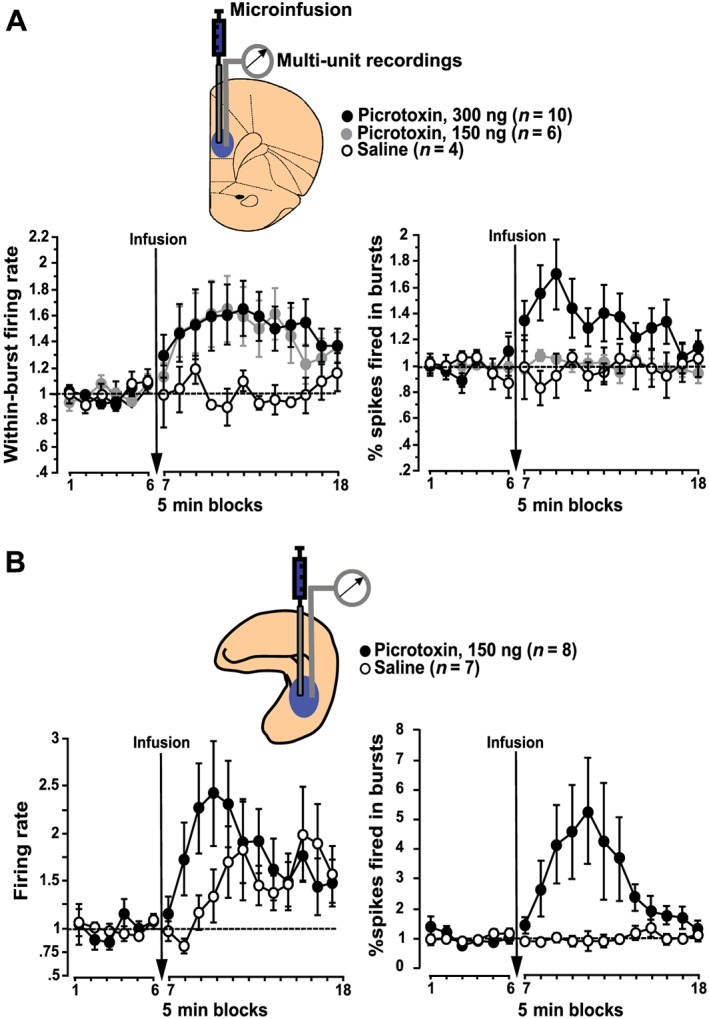
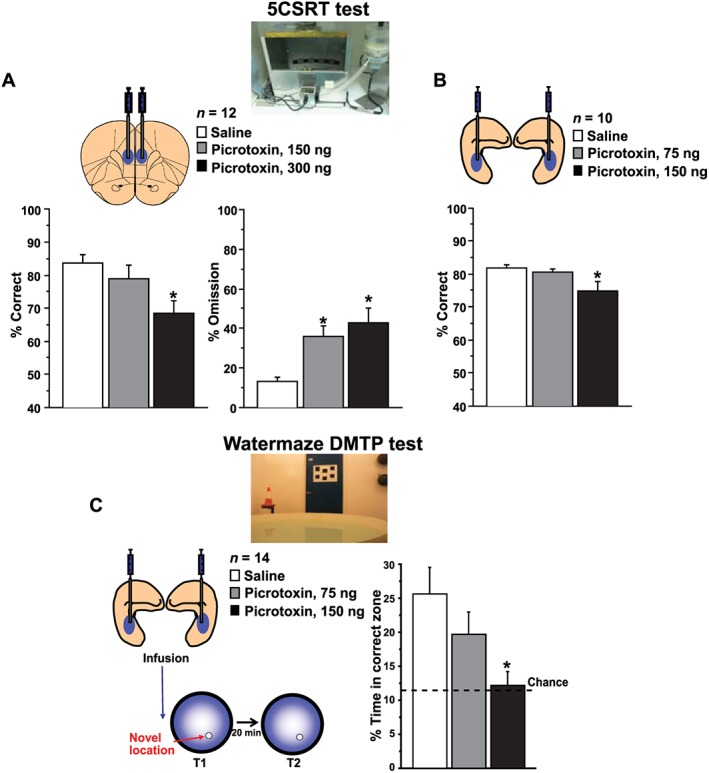
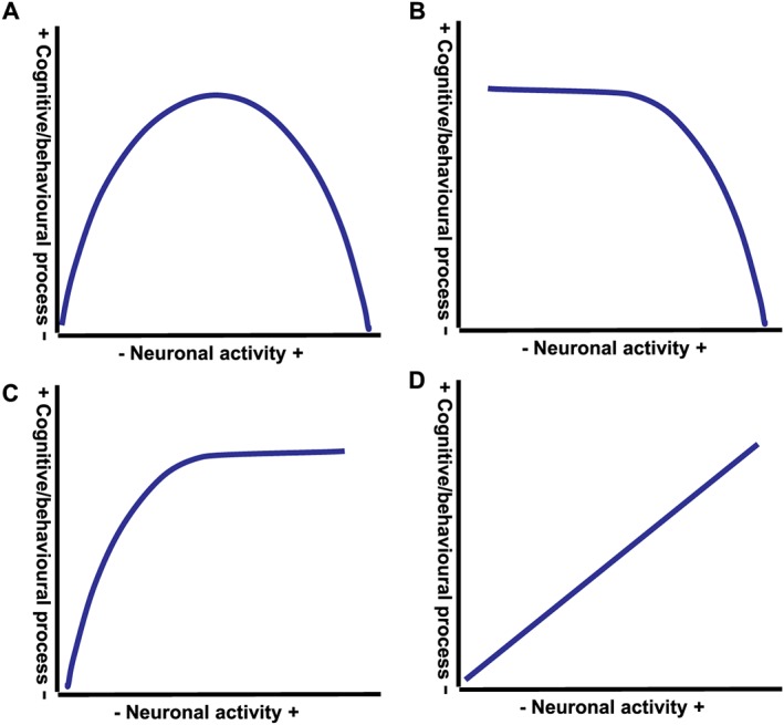
We review recent evidence concerning the significance of inhibitory GABA transmission and of neural disinhibition, that is, deficient GABA transmission, within the prefrontal cortex and the hippocampus, for clinically relevant cognitive functions. Both regions support important cognitive functions, including attention and memory, and their dysfunction has been implicated in cognitive deficits characterizing neuropsychiatric disorders. GABAergic inhibition shapes cortico-hippocampal neural activity, and, recently, prefrontal and hippocampal neural disinhibition has emerged as a pathophysiological feature of major neuropsychiatric disorders, especially schizophrenia and age-related cognitive decline. Regional neural disinhibition, disrupting spatio-temporal control of neural activity and causing aberrant drive of projections, may disrupt processing within the disinhibited region and efferent regions. Recent studies in rats showed that prefrontal and hippocampal neural disinhibition (by local GABA antagonist microinfusion) dysregulates burst firing, which has been associated with important aspects of neural information processing. Using translational tests of clinically relevant cognitive functions, these studies showed that prefrontal and hippocampal neural disinhibition disrupts regional cognitive functions (including prefrontal attention and hippocampal memory function). Moreover, hippocampal neural disinhibition disrupted attentional performance, which does not require the hippocampus but requires prefrontal-striatal circuits modulated by the hippocampus. However, some prefrontal and hippocampal functions (including inhibitory response control) are spared by regional disinhibition. We consider conceptual implications of these findings, regarding the distinct relationships of distinct cognitive functions to prefrontal and hippocampal GABA tone and neural activity. Moreover, the findings support the proposition that prefrontal and hippocampal neural disinhibition contributes to clinically relevant cognitive deficits, and we consider pharmacological strategies for ameliorating cognitive deficits by rebalancing disinhibition-induced aberrant neural activity. Linked Articles This article is part of a themed section on Pharmacology of Cognition: a Panacea for Neuropsychiatric Disease? To view the other articles in this section visit http://onlinelibrary.wiley.com/doi/10.1111/bph.v174.19/issuetoc.


Figures

Figure 1
Figure 1
Regional GABA dysfunction may disrupt regional function and the function of projection sites. Deficient function of inhibitory GABA interneurons within the prefrontal cortex or hippocampus disrupts the spatio‐temporal control of excitatory glutamatergic neurons within these regions, causing aberrant firing of these neurons (1), which may disrupt the cognitive functions normally mediated by these regions (2). In addition, such aberrant firing may cause aberrant drive of neurons in projection sites, which may disrupt the functions normally mediated by these projection sites (3).
Figure 2
Figure 2
Neural disinhibition in prefrontal cortex and hippocampus enhances burst firing of neurons within these regions. Following local microinfusions of the GABA antagonist picrotoxin into the medial prefrontal cortex (A) or hippocampus (B) of anaesthetized rats, enhanced neuronal burst firing is the most marked effect revealed by in vivo electrophysiological recordings of multi‐unit activity in the vicinity of the infusion site. (A) In the medial prefrontal cortex, regional neural disinhibition by picrotoxin primarily increases within‐burst firing rate and, at the higher picrotoxin dose, also increases the prevalence of bursts, as reflected by an increased percentage of spikes fired in bursts. (B) In the hippocampus, local picrotoxin infusion markedly increases the prevalence of bursts, as reflected, for example, by an increased percentage of spikes fired in bursts, accompanied by a comparatively moderate increase in overall firing rate. All values show multi‐unit recording data normalized to baseline (average across the six 5 min blocks before infusion) and show mean ± SEM; prefrontal data graphs are adapted from Pezze et al. (2014) and hippocampal data from McGarrity et al. (2016).
Figure 3
Figure 3
Neural disinhibition within the medial prefrontal cortex and hippocampus causes clinically relevant attentional and memory deficits. (A, B) Attentional deficits: neural disinhibition in the medial prefrontal cortex or hippocampus disrupts attentional performance on the 5CSRT task. The 5CSRT task requires rats to sustain and divide attention across a row of five apertures to detect brief light flashes occurring randomly in one of the apertures and to respond to these flashes to receive food reward. Following local microinfusions of the GABA antagonist picrotoxin into the medial prefrontal cortex (A) or hippocampus (B), rats show reduced attentional performance, as reflected by a reduced percentage of correct responses and, in case of the prefrontal disinhibition, also an increase in omitted trials. (C) Memory deficits: neural disinhibition in the hippocampus disrupts rapid place learning performance on the watermaze DMTP test. This highly hippocampus‐dependent test requires rats to learn, within one trial, the daily changing place of a hidden platform that offers escape from water, resembling the everyday task of remembering new places and routes. Following microinfusion of picrotoxin into the hippocampus, rats show reduced one‐trial place learning performance, as reflected by a marked reduction in search preference for the correct location. Prefrontal data graphs are adapted from Pezze et al. (2014) and hippocampal data graphs from McGarrity et al. (2016).
Figure 4
Figure 4
Distinct causal relationships link neural activity within medial prefrontal cortex and hippocampus to distinct cognitive and behavioural processes. Studies examining the cognitive and behavioural impact of bidirectional changes of neural activity within the medial prefrontal cortex or hippocampus, including by local infusions of GABA agonists and antagonists, show that neural activity within these regions can be linked to distinct cognitive and behavioural processes by distinct causal relationships. (A) Inverted U‐shaped relationship, with cognitive or behavioural process requiring a balanced level of neural activity and both too little and too much neural activity causing impairments; for example, the relationship between prefrontal neural activity and attentional performance and between hippocampal neural activity and rapid place learning performance. (B) Cognitive or behavioural process is not affected by reductions in neural activity but impaired by increases in neural activity; for example, the relationship between hippocampal neural activity and attentional performance. (C) Cognitive or behavioural process can be sustained as long as neural activity is above a minimal level; for example, the relationship between hippocampal and prefrontal neural activity and response control. (D) Monotonic positive relationship between neural activity and cognitive or behavioural process, with decreases in neural activity reducing the process and increases in neural activity increasing the process; for example, the relationship between hippocampal and prefrontal neural activity and locomotor activity.

Similar articles

Cited by

References

    1. Alexander SPH, Peters JA, Kelly E, Marrion N, Benson HE, Faccenda E et al. (2015a). The Concise Guide to PHARMACOLOGY 2015/16: Ligand‐gated ion channels. Br J Pharmacol 172: 5870–5903. - PMC - PubMed
    1. Alexander SPH, Davenport AP, Kelly E, Marrion N, Peters JA, Benson HE et al. (2015b). The Concise Guide to PHARMACOLOGY 2015/16: G protein‐coupled receptors. Br J Pharmacol 172: 5744–5869. - PMC - PubMed
    1. Andrews‐Zwilling Y, Gillespie AK, Kravitz AV, Nelson AB, Devidze N, Lo I et al. (2012). Hilar GABAergic interneuron activity controls spatial learning and memory retrieval. PLoS One 7: e40555. - PMC - PubMed
    1. Anticevic A, Corlett PR, Cole MW, Savic A, Gancsos M, Tang Y et al. (2015). N‐methyl‐D‐aspartate receptor antagonist effects on prefrontal cortical connectivity better model early than chronic schizophrenia. Biol Psychiatry 77: 569–580. - PubMed
    1. Anticevic A, Gancsos M, Murray JD, Repovs G, Driesen NR, Ennis DJ et al. (2012). NMDA receptor function in large‐scale anticorrelated neural systems with implications for cognition and schizophrenia. Proc Nat Acad Sci 109: 16720–16725. - PMC - PubMed

Substances

LinkOut - more resources