Review
. 2017 Jun;10(2):399-408.
doi: 10.1016/j.path.2017.01.013. Epub 2017 Mar 14.

Merkel Cell Carcinoma

Affiliations
Review

Merkel Cell Carcinoma

Melissa Pulitzer. Surg Pathol Clin. 2017 Jun.

Abstract

Merkel cell carcinoma (MCC) encompasses neuroendocrine carcinomas primary to skin and occurs most commonly in association with clonally integrated Merkel cell polyomavirus with related retinoblastoma protein sequestration or in association with UV radiation-induced alterations involving the TP53 gene and mutations, heterozygous deletion, and hypermethylation of the Retinoblastoma gene. Molecular genetic signatures may provide therapeutic guidance. Morphologic features, although patterned, are associated with predictable diagnostic pitfalls, usually resolvable by immunohistochemistry. Therapeutic options for MCC, traditionally limited to surgical intervention and later chemotherapy and radiation, are growing, given promising early results of immunotherapeutic regimens.

Keywords: Merkel cell; Neuroendocrine; Polyomavirus; UV.


Figures

Fig. 1
Fig. 1
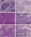
(A). Cytomorphology of MCC showing round nuclei, scant cytoplasm, salt-and-pepper chromatin, indistinct nucleoli [Hematoxylin-eosin, original magnification ×200]. (B) Combined MCC; a small cell component demonstrates small round nuclei with crush artifact whereas a second more epitheloid population is punctuated by large keratinizing squamous cells [Hematoxylin-eosin, original magnification ×400]. (C) Intraepidermal pagetoid extension of MCC overlying a dermal tumor nodule (focally seen in left lower corner) [Hematoxylin-eosin, original magnification ×400]. (D) MCC in situ arising within severely dysplastic squamous epithelium/SCC in situ of epithelium, associated with invasive NEC in adjacent dermis [Hematoxylin-eosin, original magnification ×400]. (E) Low-power photomicrograph showing nodular circumscription of a dermal tumor [Hematoxylin-eosin, original magnification ×40]. (F) Intralymphatic tumor emboli of MCC within ectatic deep dermal lymphatics [Hematoxylin-eosin, original magnification ×400].
Fig. 2
Fig. 2
(A) CK20 stain in a combined tumor shows a varied pattern of labeling, including patchy but diffuse perinuclear dots as well as areas of membranous accentuation [Hematoxylin-eosin, original magnification ×400]. Squamous cell shows strong cytoplasmic labeling as well [Hematoxylin-eosin, original magnification ×200]. (B) Polyomavirus immunohistochemical stain diffusely labels tumor nuclei [Hematoxylin-eosin, original magnification ×400].
Fig. 3
Fig. 3
Differential diagnostic considerations of MCC. (A) Islands of small round blue cells with mucinous tumor-stroma retraction in MCC mimicking BCC [Hematoxylin-eosin, original magnification ×400]. (B) Extensive intraepidermal and pagetoid spread in MCC mimicking melanoma in situ [Hematoxylin-eosin, original magnification ×200]. (C) Pleomorphism (architecturally and cytologically) characteristic of a metastatic NEC, in a tumor derived from the lung [Hematoxylin-eosin, original magnification ×400]. (D) WDNET/carcinoid metastatic to the skin shows organized, organoid architecture and lacks high grade nuclear features [Hematoxylin-eosin, original magnification ×400]. (E) In situ component of MCC; some intraepidermal tumor cells appear to have bubbly cytoplasm and an intrafollicular localization reminiscent of sebaceous carcinoma [Hematoxylin-eosin, original magnification ×400]. (F) Combined MCC/SCC; immunohistochemistry would be needed to distinguish from poorly differentiated SCC [Hematoxylin-eosin, original magnification ×100].
Fig. 4
Fig. 4
Diagnostic algorithm for MCC. H&E, hematoxylin-eosin stain.

Similar articles

Cited by

References

    1. Bichakjian CK, Lowe L, Lao CD, et al. Merkel cell carcinoma: critical review with guidelines for multi-disciplinary management. Cancer. 2007;110:1–12. - PubMed
    1. Agelli M, Clegg LX. Epidemiology of primary Merkel cell carcinoma in the United States. J Am Acad Dermatol. 2003;49:832–41. - PubMed
    1. Sihto H, Kukko H, Koljonen V, et al. Merkel cell polyomavirus infection, large T antigen, retinoblastoma protein and outcome in Merkel cell carcinoma. Clin Cancer Res. 2011;17:4806–13. - PubMed
    1. Suarez AL, Louis P, Kitts J, et al. Clinical and dermoscopic features of combined cutaneous squamous cell carcinoma (SCC)/neuroendocrine [Merkel cell] carcinoma (MCC) J Am Acad Dermatol. 2015;73:968–75. - PMC - PubMed
    1. Bichakjian CK, Hghiem P, Johnson T, et al. Merkel cell carcinoma. New York: Springer; 2016.

MeSH terms

Substances