

Chronic tinnitus and unipolar brush cell alterations in the cerebellum and dorsal cochlear nucleus
- PMID: 28478300
- PMCID: PMC5518479
- DOI: 10.1016/j.heares.2017.04.016
Chronic tinnitus and unipolar brush cell alterations in the cerebellum and dorsal cochlear nucleus
Abstract
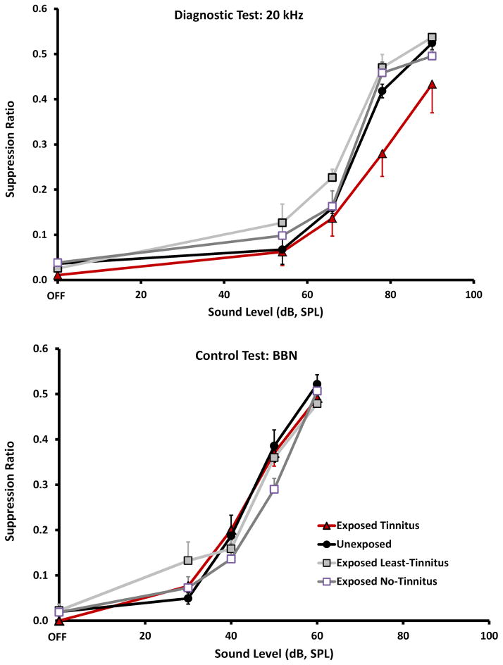
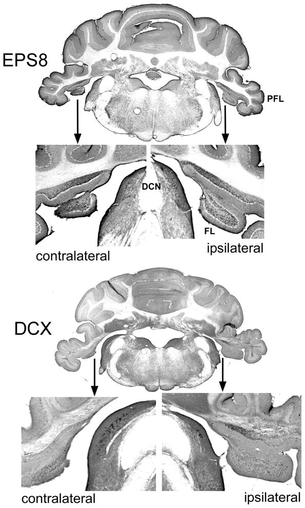
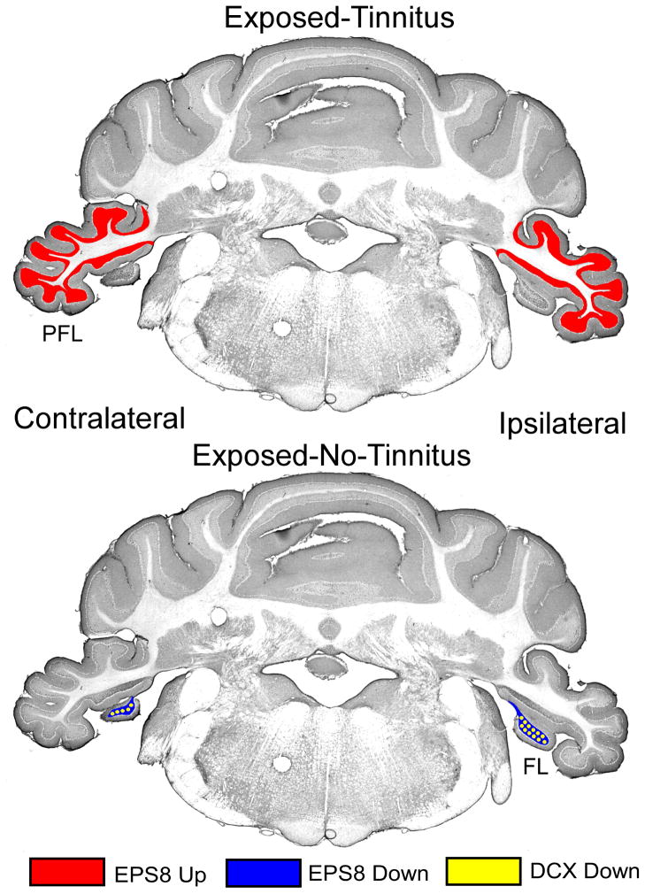
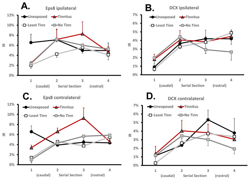
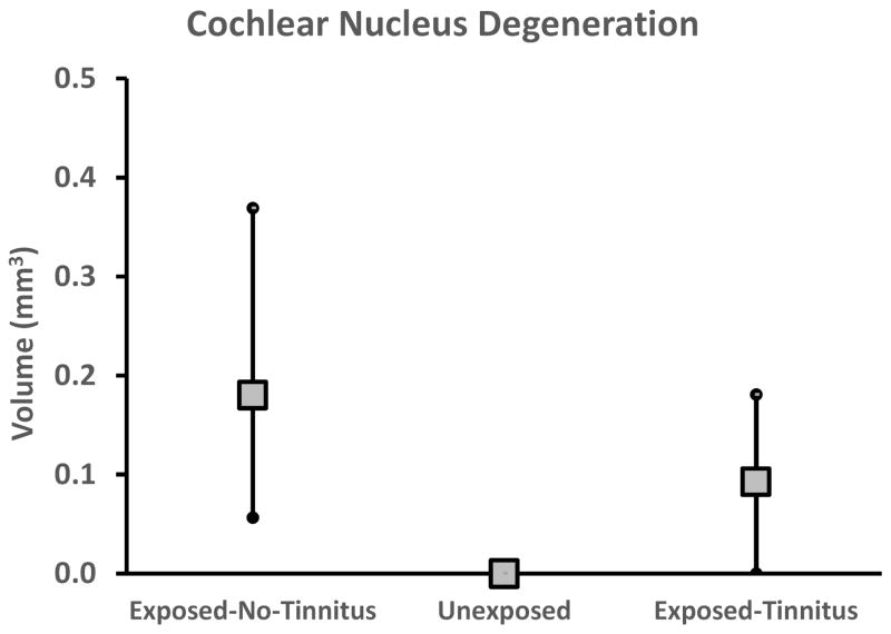
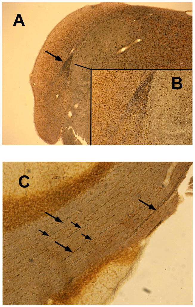
Animal model research has shown that the central features of tinnitus, the perception of sound without an acoustic correlate, include elevated spontaneous and stimulus-driven activity, enhanced burst-mode firing, decreased variance of inter-spike intervals, and distortion of tonotopic frequency representation. Less well documented are cell-specific correlates of tinnitus. Unipolar brush cell (UBC) alterations in animals with psychophysical evidence of tinnitus has recently been reported. UBCs are glutamatergic interneurons that appear to function as local-circuit signal amplifiers. UBCs are abundant in the dorsal cochlear nucleus (DCN) and very abundant in the flocculus (FL) and paraflocculus (PFL) of the cerebellum. In the present research, two indicators of UBC structure and function were examined: Doublecortin (DCX) and epidermal growth factor receptor substrate 8 (Eps8). DCX is a protein that binds to microtubules where it can modify their assembly and growth. Eps8 is a cell-surface tyrosine kinase receptor mediating the response to epidermal growth factor; it appears to have a role in actin polymerization as well as cytoskeletal protein interactions. Both functions could contribute to synaptic remodeling. In the present research UBC Eps8 and DCX immunoreactivity (IR) were determined in 4 groups of rats distinguished by their exposure to high-level sound and psychophysical performance: Unexposed, exposed to high-level sound with behavioral evidence of tinnitus, and two exposed groups without behavioral evidence of tinnitus. Compared to unexposed controls, exposed animals with tinnitus had Eps8 IR elevated in their PFL; other structures were not affected, nor was DCX IR affected. This was interpreted as UBC upregulation in animals with tinnitus. Exposure that failed to produce tinnitus did not increase either Eps8 or DCX IR. Rather Eps8 IR was decreased in the FL and DCN of one subgroup (Least-Tinnitus), while DCX IR decreased in the FL of the other subgroup (No-Tinnitus). Neuron degeneration was also documented in the cochlear nucleus and PFL of exposed animals, both with and without tinnitus. Degeneration was not found in unexposed animals. Implications for tinnitus neuropathy are discussed in the context of synaptic remodeling and cerebellar sensory modulation.
Keywords: Animal model; Auditory afferent degeneration; Cerebellum; Doublecortin; Eps8; Tinnitus; Unipolar brush cell.
Copyright © 2017 Elsevier B.V. All rights reserved.
Figures

References
-
- Azizi SA, Burne RA, Woodward DJ. The auditory corticopontocerebellar projection in the rat: inputs to the paraflocculus and midvermis. An anatomical and physiological study. Exp Brain Res. 1985;59:36–49. - PubMed
-
- Bauer CA, Ciobanu L, Brozoski TJ. Central neural activity in rats with tinnitus evaluated with MEMRI. International Society for Magnetic Resonance Imaging; Seattle, Washington: 2006. - PubMed
-
- Bauer CA, Brozoski TJ, Myers K. Primary afferent dendrite degeneration as a cause of tinnitus. J Neurosci Res. 2007;85:1489–98. - PubMed
MeSH terms
Substances
Grants and funding
LinkOut - more resources
Full Text Sources
Other Literature Sources
Medical
Research Materials
Miscellaneous
