. 2017 Aug;47(11):2017-2027.
doi: 10.1017/S0033291717000460. Epub 2017 May 8.

Veterans with post-traumatic stress disorder exhibit altered emotional processing and attentional control during an emotional Stroop task

Affiliations

Veterans with post-traumatic stress disorder exhibit altered emotional processing and attentional control during an emotional Stroop task

M M Khanna et al. Psychol Med. 2017 Aug.

Abstract

Background: Post-traumatic stress disorder (PTSD) is often associated with attention allocation and emotional regulation difficulties, but the brain dynamics underlying these deficits are unknown. The emotional Stroop task (EST) is an ideal means to monitor these difficulties, because participants are asked to attend to non-emotional aspects of the stimuli. In this study, we used magnetoencephalography (MEG) and the EST to monitor attention allocation and emotional regulation during the processing of emotionally charged stimuli in combat veterans with and without PTSD.

Method: A total of 31 veterans with PTSD and 20 without PTSD performed the EST during MEG. Three categories of stimuli were used, including combat-related, generally threatening and neutral words. MEG data were imaged in the time-frequency domain and the network dynamics were probed for differences in processing threatening and non-threatening words.

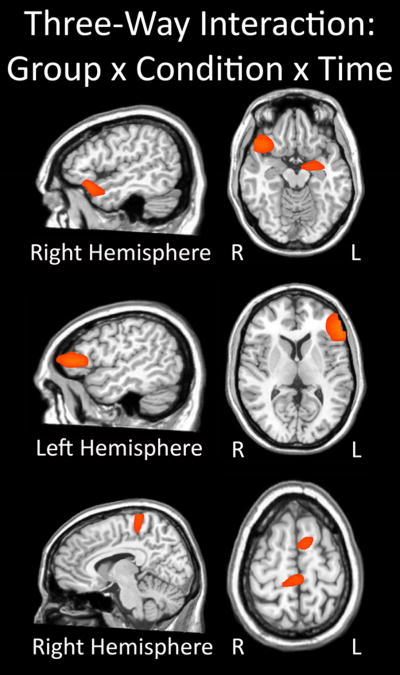
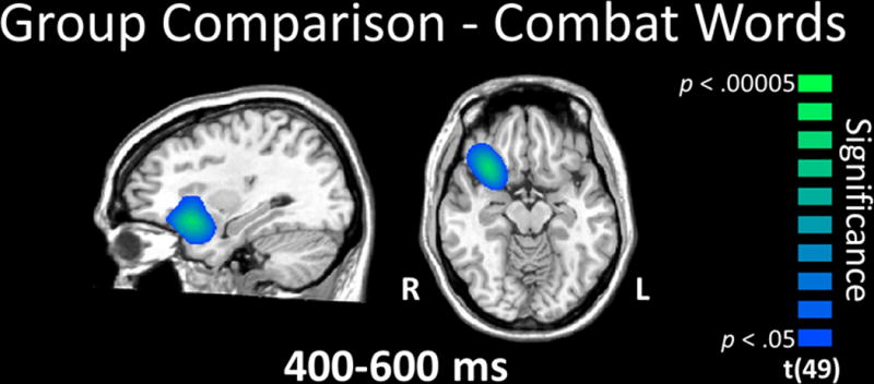
Results: Behaviorally, veterans with PTSD were significantly slower in responding to combat-related relative to neutral and generally threatening words. Veterans without PTSD exhibited no significant differences in responding to the three different word types. Neurophysiologically, we found a significant three-way interaction between group, word type and time period across multiple brain regions. Follow-up testing indicated stronger theta-frequency (4-8 Hz) responses in the right ventral prefrontal (0.4-0.8 s) and superior temporal cortices (0.6-0.8 s) of veterans without PTSD compared with those with PTSD during the processing of combat-related words.

Conclusions: Our data indicated that veterans with PTSD exhibited deficits in attention allocation and emotional regulation when processing trauma cues, while those without PTSD were able to regulate emotion by directing attention away from threat.

Keywords: Attention allocation; emotional Stroop task; emotional regulation; oscillation; post-traumatic stress disorder.


Conflict of interest statement

Conflict of interest: None

Figures

Figure 1
Figure 1
Single trial design and overall layout of the emotional Stroop Task
Figure 2
Figure 2
Three-way interaction of group-by-condition-by-time. All three panels show brain regions exhibiting a significant three-way interaction effect (p < 0.05). Significant neural areas included the right inferior frontal, ventral frontal, and superior temporal regions, as well as the left amygdala (top panel). In addition, the three-way interaction was significant in the left inferior frontal gyrus and dorsolateral prefrontal cortex region (middle panel), as well as the cingulate cortices and superior frontal (bottom panel). All three panels have been thresholded at (p < 0.05) and are displayed in radiological convention. The three-way interaction effect consisted of group (2 levels; veterans with and without PTSD), condition (3 levels; Combat-related threat words, general threat words, and neutral words), and time (4 levels; 0.0 – 0.2 s, 0.2 – 0.4 s, 0.4 – 0.6 s, and 0.6 – 0.8 s).
Figure 3
Figure 3
Group comparison for combat-related words. Veterans without PTSD exhibited significantly stronger theta activity in right ventral prefrontal cortex (PFC) during the 0.4 - 0.6 s time window relative to veterans with PTSD (p < 0.05, corrected). This group difference in the ventral PFC was sustained into the 0.6 - 0.8 s time window, and this latter window also included the right temporal cortices (not shown). All images are displayed by radiological convention (R=L) and have been thresholded at (p < 0.05, corrected).
Figure 4
Figure 4
Word list (conditional) effects in each group. (Top panel) General threat compared to neutral words. During early processing (0.2 – 0.4 s), veterans without PTSD exhibited significantly stronger theta activity in the left dorsolateral prefrontal cortices during general threat compared to neutral words, and this activity spread to the left inferior frontal, superior temporal, and ventral PFC, as well as the right supramarginal gyrus later in the time course. In contrast, veterans with PTSD exhibited significant differences between general threat and neutral words during only the 0.6 - 0.8 s time window, and these differences reflected greater theta for the general threat words in the left hippocampus and amygdala, as well as less theta within the left medial prefrontal cortex. (Bottom left panel) Combat-related threat compared to neutral words. Veterans without PTSD exhibited greater theta in left fronto-temporal cortices during combat-related words relative to neutral words in the 0.4 - 0.6 s time window. Slightly later, veterans without PTSD had significantly stronger theta activity in the right ventral PFC and medial temporal areas, including the amygdala, in response to combat-related words. Veterans with PTSD had significantly stronger theta activity during neutral relative to combat word processing in the parieto-occipital (0.4 – 0.6 s) and cingulate cortices (0.6 – 0.8 s). (Bottom right panel) Combat-related compared to general threat words. There were no significant differences between combat and general threat words in veterans without PTSD in any time window. In the 0.4 – 0.6 s time bin, theta activity was weaker for combat-related compared to general-threat words in the right ventral PFC and medial temporal structures (hippocampus/amygdala), extending into more lateral temporal cortices, as well as the cingulate cortices. All images are displayed following radiological convention (R=L) and have been thresholded at (p < 0.05, corrected).

Similar articles

Cited by

References

    1. Anders SL, Peterson CK, James LM, Engdahl B, Leuthold AC, Georgopoulos AP. Neural communication in posttraumatic growth. Experimental Brain Research. 2015;233(7):2013–2020. - PubMed
    1. Aupperle RL, Melrose AJ, Stein MB, Paulus MP. Executive function and PTSD: Disengaging from trauma. Neuropharmacology. 2012;62:686–694. - PMC - PubMed
    1. Badura-Brack AS, Naim R, Ryan TJ, Levy O, Abend R, Khanna MM, McDermott TJ, Pine DS, Bar-Haim Y. Effect of attention training on attention bias variability and PTSD symptoms: Randomized controlled trials in Israeli and U.S. combat veterans. American Journal of Psychiatry. 2015;172(12):1233–1241. - PMC - PubMed
    1. Balota DA, Yap MJ, Cortese MJ, Hutchison KA, Kessler B, Loftis B, Neely JH, Nelson DL, Simpson GB, Treiman R. The English Lexicon Project. Behavior Research Methods. 2007;39:445–459. - PubMed
    1. Bar-Haim Y. Research Review: attention bias modification (ABM): a novel treatment for anxiety disorders. Journal of Child Psychology and Psychiatry. 2010;51:859–870. - PubMed