

Epithelioid hemangioendothelioma of the craniocervical junction; case report and review
- PMID: 28478911
- PMCID: PMC6197577
- DOI: 10.1016/j.aott.2017.03.012
Epithelioid hemangioendothelioma of the craniocervical junction; case report and review
Abstract
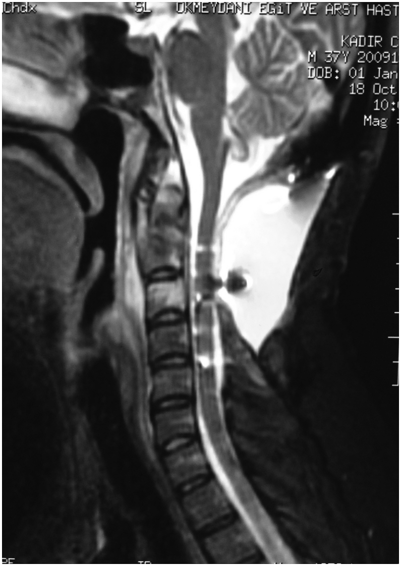
Epithelioid hemangioendotheliomas are uncommon vascular neoplasms and their spinal location is even rarer. We report clinical course of a 31-year-old man with an epithelioid hemangioendothelioma at the cranio-cervical junction. A cervical magnetic resonance imaging revealed tumor that caused posterior cervical cord compression. C1,2,3 total laminectomy and surgical excision of the tumor was performed. Postoperative external beam radiation was performed on the surgical field especially around the right vertebral artery. At 2-year follow-up there was no neurological deficit and no tumor recurrence.
Keywords: Craniocervical junction; Epithelioid hemangioendothelioma; Surgical excision.
Copyright © 2017 Turkish Association of Orthopaedics and Traumatology. Production and hosting by Elsevier B.V. All rights reserved.
Figures


References
-
- Christodoulou A., Symeonidis P.D., Kapoutsis D., Iordanidis F. Primary epithelioid hemangioendothelioma of the lumbar spine. Spine. 2008;8(2):385–390. - PubMed
-
- Brennan J.W., Midha R., Ang L.C., Perez-Ordonez B. Epithelioid hemangioendothelioma of the spine presenting as cervical myelopathy: case report. Neurosurgery. 2001;48(5):1166–1169. - PubMed
-
- Gokhan G.A., Akyuz M., Gurer I.E., Tuncer R. Epithelioid hemangioendothelioma derived from the spine region: case report and review of the literature. Wien Klin Wochenschr. 2006;118(11–12):358–361. - PubMed
-
- Aquilina K., Lim C., Kamel M.H., Marks C.J., O'Sullivan M.G., Keohane C. Epithelioid hemangioendothelioma of the spine. Report of two cases. J Neurosurg Spine. 2005;3(5):393–399. - PubMed
Publication types
MeSH terms
LinkOut - more resources
Full Text Sources
Other Literature Sources
