. 2017 Jun 6;8(6):1525-1533.
doi: 10.1016/j.stemcr.2017.04.004. Epub 2017 May 4.

Comprehensive Monosynaptic Rabies Virus Mapping of Host Connectivity with Neural Progenitor Grafts after Spinal Cord Injury

Affiliations

Comprehensive Monosynaptic Rabies Virus Mapping of Host Connectivity with Neural Progenitor Grafts after Spinal Cord Injury

Andrew F Adler et al. Stem Cell Reports. .

Abstract

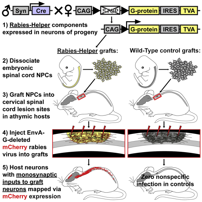
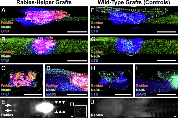
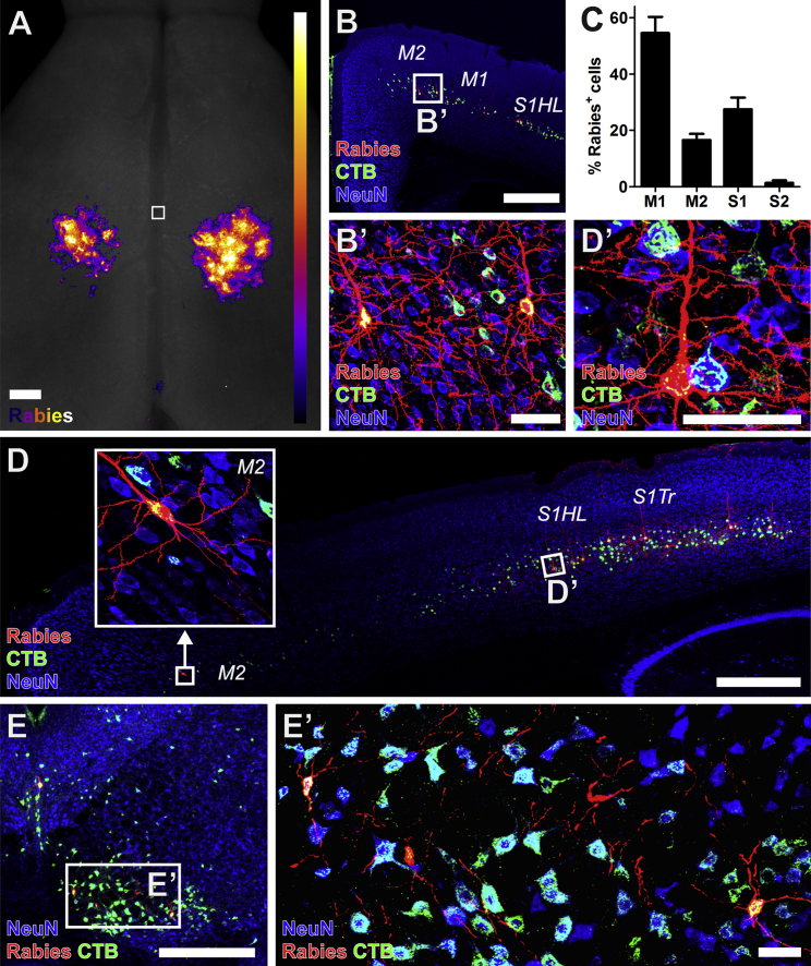
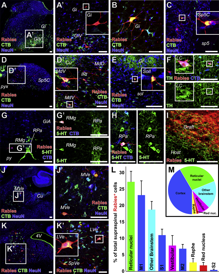
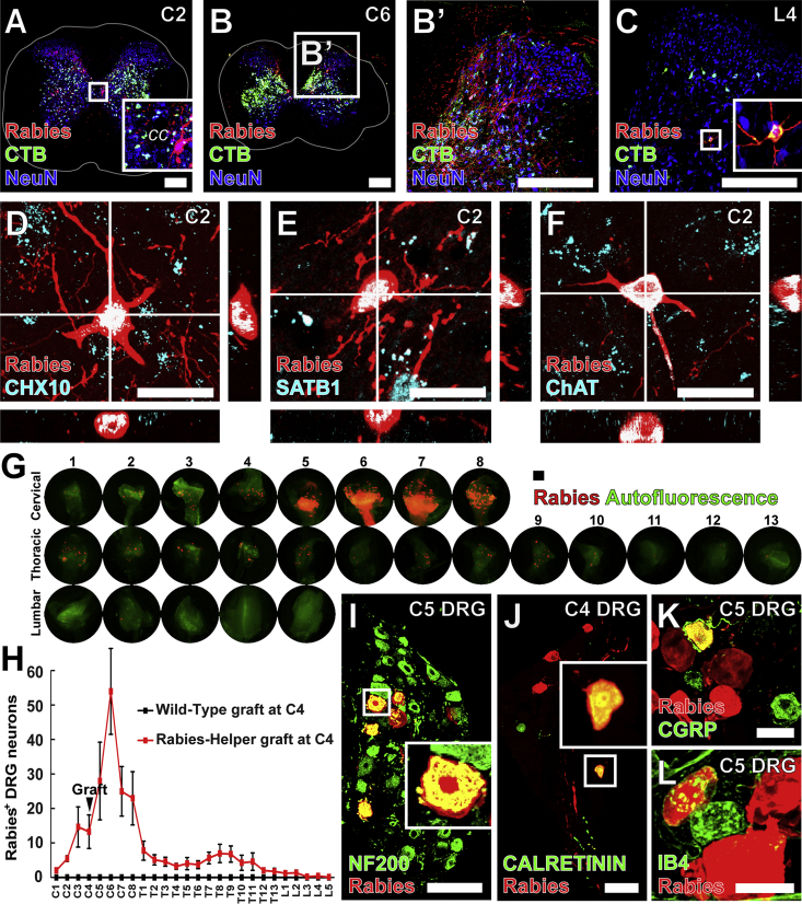
Neural progenitor cells grafted to sites of spinal cord injury have supported electrophysiological and functional recovery in several studies. Mechanisms associated with graft-related improvements in outcome appear dependent on functional synaptic integration of graft and host systems, although the extent and diversity of synaptic integration of grafts with hosts are unknown. Using transgenic mouse spinal neural progenitor cell grafts expressing the TVA and G-protein components of the modified rabies virus system, we initiated monosynaptic tracing strictly from graft neurons placed in sites of cervical spinal cord injury. We find that graft neurons receive synaptic inputs from virtually every known host system that normally innervates the spinal cord, including numerous cortical, brainstem, spinal cord, and dorsal root ganglia inputs. Thus, implanted neural progenitor cells receive an extensive range of host neural inputs to the injury site, potentially enabling functional restoration across multiple systems.

Keywords: cell therapy; grafting; monosynaptic; neural progenitor cells; neural regeneration; rabies; spinal cord injury; transsynaptic.


Figures

None
Graphical abstract
Figure 1
Figure 1
Monosynaptically Restricted Rabies Infection Initiated Exclusively from Rabies-Helper Spinal NPC Grafts after SCI Grafts of either (A–E) Rabies-Helper-expressing NPC grafts or (F–J) Wild-Type grafts lacking rabies-helper components. (A) Sagittal view near the central canal of a Rabies-Helper NPC graft in a C4 spinal cord lesion site 3 months after lesion/grafting and 1 week after EnvA-SADΔG-mCherry rabies virus (red) injection into graft. CTB (blue) co-injected with rabies diffuses beyond the graft, whereas mCherry expression is strictly initiated from graft neurons. NeuN, green. Rostral is to the left, caudal to the right. (B) Sagittal view through a more lateral section of a Rabies-Helper NPC graft, showing the extent of injury to host gray matter. (C) Transverse view of a Rabies-Helper graft in a second animal, showing that the lesion extends to involve some of the lateral and ventral white matter. (D) Sagittal view of MAP2 (blue) and NeuN (green) expression in graft neurons. (E) Gross horizontal view of brain/brainstem/spinal cord preparation, showing intense expression of Rabies-mCherry under fluorescent illumination in the graft at the lesion site (white, center of image) and retrograde transsynaptic expression of Rabies-mCherry in the cortex (arrows) and in several dorsal root ganglia (arrowheads). A sample DRG is shown at higher magnification in the inset. (F–J) In animals that received Wild-Type grafts followed by injections of SADΔG-mCherry rabies virus and CTB, there is not a single cell expressing Rabies-mCherry in the graft (F–I) or at remote host locations (J). Scale bars, 1 mm. Dotted lines denote graft-host borders. Solid lines denote the extent of white matter in transverse sections. See also Figure S1.
Figure 2
Figure 2
Host Cortical and Rubral Neurons Make Monosynaptic Contact with Graft Neurons (A) Heatmap of the location of Rabies-mCherry+ corticospinal neurons making monosynaptic contact onto Rabies-Helper spinal NPC graft-derived neurons (aggregate of n = 8 mice). Horizontal view. Square denotes bregma, with rostral at the top of the image. Most of the Rabies-mCherry signals from the apical dendrites of corticospinal neurons are located caudal to the bregma, in the caudal forelimb and hindlimb regions. (B and C) Rabies-mCherry+ (red) cortical neurons making synaptic contacts onto graft neurons co-labeled with CTB injected into graft sites (green), and were (C) found most frequently in the primary motor cortex (M1; mean of all Rabies-Helper grafted mice cut coronally, ± SEM, n = 5). NeuN, blue. (D) Rabies-mCherry+ cortical neurons making synaptic contacts onto graft neurons were also found in the secondary motor cortex (M2, boxed detail) in the rostral forelimb area, and hindlimb and trunk primary somatosensory cortex (S1HL, S1Tr). (E) Rabies-mCherry+ neurons were also present in the red nucleus. 30-μm-thick sections. Scale bars, 500 μm in (A, B, D, and E) and 50 μm in (B′, D′, and E’). See also Figures S2 and S4.
Figure 3
Figure 3
Host Brainstem Neurons Make Monosynaptic Contact with Graft Neurons (A–H) Host Rabies-mCherry+ (red) neurons making monosynaptic contact onto Rabies-Helper graft neurons co-labeled with CTB injected into graft sites were found in (A and B) the gigantocellular (Gi) and gigantocellular, ventral part (GiV) reticular nuclei, (C) the caudal part of the spinal trigeminal nucleus (Sp5C) adjacent to the spinal trigeminal tract (sp5), (D) the ventral/intermediate/dorsal parts of the medullary reticular nucleus (MdV/IRt/MdD) adjacent to the cuneate nucleus (Cu) and pyramidal decussation (pyx), and (E) the interstitial part of the nucleus of the solitary tract (SolI) adjacent to the solitary tract (sol) and gracile nucleus (Gr). Host mCherry+CTB+ neurons making monosynaptic contact with graft neurons were also co-labeled with tyrosine hydroxylase (TH, green) in the locus coeruleus (LC) (F), and in the raphe magnus (RMg) (G), and the raphe pallidus (RPa) (H) co-labeled with serotonin (5-HT, green) adjacent to the pyramidal tracts (py) and alpha part of the gigantocellular reticular nucleus (GiA). (I) Serotonergic host axons penetrated grafts. (J and K) Host mCherry+CTB+ neurons making monosynaptic contact with graft neurons were also found in the medial vestibular nucleus (MVe) adjacent to cuneate nucleus (Cu) (J), and in the lateral and spinal vestibular nuclei (LVe, SpVe) adjacent to the fourth ventricle (4V) (K). (L) Among all supraspinal Rabies-mCherry+ neurons, those found in reticular and corticospinal nuclei were the most common (mean ± SEM of all Rabies-Helper-grafted mice cut transversely, n = 5). (M) Pie chart representation of data in (L). Insets depict detail of boxed regions. Transverse sections, except sagittal in (J and J’). 30-μm-thick sections. Scale bars, 100 μm. See also Figure S3.
Figure 4
Figure 4
Host Spinal and Dorsal Root Ganglia Neurons Make Monosynaptic Contact with Graft-Derived Neurons (A–C) Host Rabies-mCherry+ (red) neurons making monosynaptic contact with Rabies-Helper graft neurons co-labeled with CTB (green) injected into graft sites were found in the cervical spinal cord rostral (A) and caudal (B) to graft sites, as well as less frequently at lumbar levels (transverse sections) (C). White lines denote the extent of white matter. (D and E) At C2, Rabies-mCherry+CHX10+ (D), and Rabies-mCherry+SATB1+ neurons (E) were present in the intermediate gray matter where premotor CHX10+ V2a and SATB1/2+ interneurons are located. (F) Rabies-mCherry+ChAT+ neurons were also found adjacent to the central canal (cc), where premotor V0c ChAT+ neurons are located. Transverse sections. (G) Rabies-mCherry+ DRG neurons making monosynaptic contact with graft neurons were found at cervical, thoracic, and lumbar levels, both rostral and caudal to the injury and graft site. (H) Most labeled DRG neurons were found at cervical levels immediately caudal to the graft site (mean ± SEM, n = 8 Rabies-Helper grafted mice, n = 3 Wild-Type grafted mice). (I–L) Some large-diameter Rabies-mCherry+ DRG neurons expressed (I) neurofilament 200 (NF200) or (J) CALRETININ, and some small-diameter Rabies-mCherry+ DRG neurons (K) expressed calcitonin gene-related peptide (CGRP), or (L) bound isolectin B4 (IB4), marking sensory neurons responsive to (I and J) touch and (K and L) noxious stimuli. Insets depict detail of boxed regions. 30-μm-thick sections for (A–F), 20-μm-thick sections for (I–K). Scale bars, 250 μm in (A–C), 20 μm in (D–F), 100 μm in (G, I, and J), and 30 μm in (K and L). See also Figure S3.

References

    1. Azim E., Jiang J., Alstermark B., Jessell T.M. Skilled reaching relies on a V2a propriospinal internal copy circuit. Nature. 2014;508:357–363. - PMC - PubMed
    1. Basaldella E., Takeoka A., Sigrist M., Arber S. Multisensory signaling shapes vestibulo-motor circuit specificity. Cell. 2015;163:301–312. - PubMed
    1. Bonner J.F., Connors T.M., Silverman W.F., Kowalski D.P., Lemay M.A., Fischer I. Grafted neural progenitors integrate and restore synaptic connectivity across the injured spinal cord. J. Neurosci. 2011;31:4675–4686. - PMC - PubMed
    1. Bourane S., Grossmann K.S., Britz O., Dalet A., Del Barrio M.G., Stam F.J., Garcia-Campmany L., Koch S., Goulding M. Identification of a spinal circuit for light touch and fine motor control. Cell. 2015;160:503–515. - PMC - PubMed
    1. Deumens R., Koopmans G.C., Joosten E.A. Regeneration of descending axon tracts after spinal cord injury. Prog. Neurobiol. 2005;77:57–89. - PubMed

Publication types

MeSH terms