

High-resolution magnetic resonance imaging reveals nuclei of the human amygdala: manual segmentation to automatic atlas
- PMID: 28479476
- PMCID: PMC5557007
- DOI: 10.1016/j.neuroimage.2017.04.046
High-resolution magnetic resonance imaging reveals nuclei of the human amygdala: manual segmentation to automatic atlas
Abstract
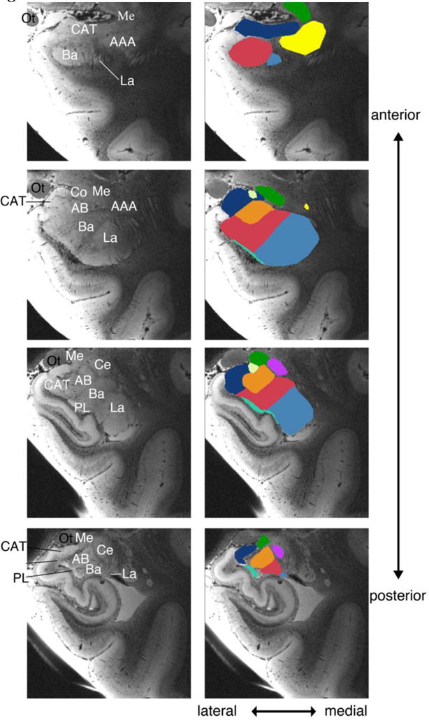
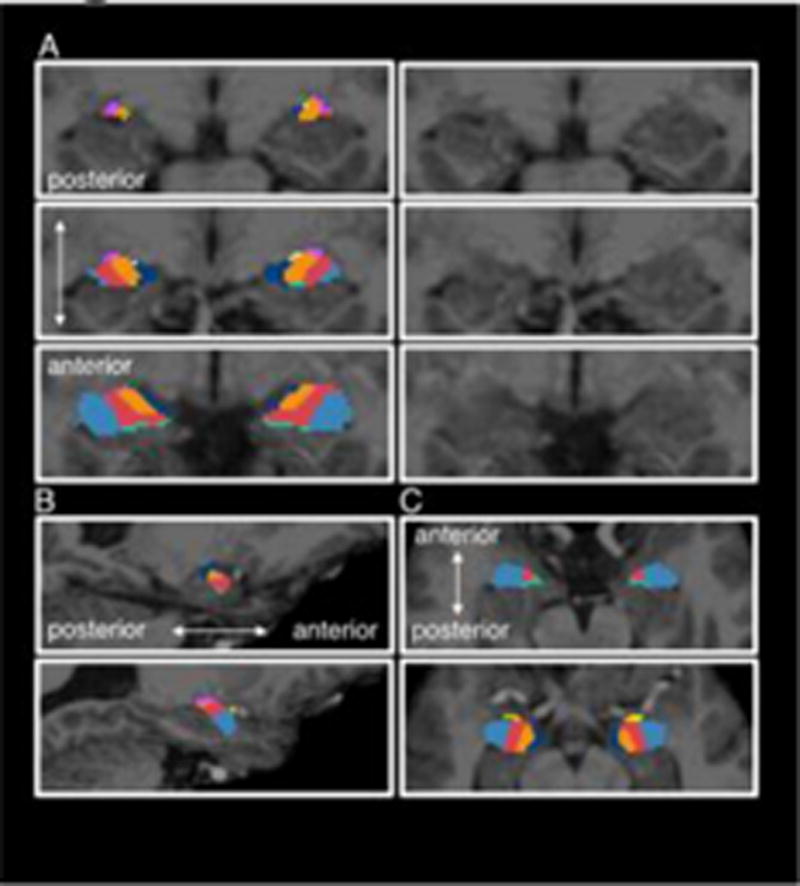
The amygdala is composed of multiple nuclei with unique functions and connections in the limbic system and to the rest of the brain. However, standard in vivo neuroimaging tools to automatically delineate the amygdala into its multiple nuclei are still rare. By scanning postmortem specimens at high resolution (100-150µm) at 7T field strength (n = 10), we were able to visualize and label nine amygdala nuclei (anterior amygdaloid, cortico-amygdaloid transition area; basal, lateral, accessory basal, central, cortical medial, paralaminar nuclei). We created an atlas from these labels using a recently developed atlas building algorithm based on Bayesian inference. This atlas, which will be released as part of FreeSurfer, can be used to automatically segment nine amygdala nuclei from a standard resolution structural MR image. We applied this atlas to two publicly available datasets (ADNI and ABIDE) with standard resolution T1 data, used individual volumetric data of the amygdala nuclei as the measure and found that our atlas i) discriminates between Alzheimer's disease participants and age-matched control participants with 84% accuracy (AUC=0.915), and ii) discriminates between individuals with autism and age-, sex- and IQ-matched neurotypically developed control participants with 59.5% accuracy (AUC=0.59). For both datasets, the new ex vivo atlas significantly outperformed (all p < .05) estimations of the whole amygdala derived from the segmentation in FreeSurfer 5.1 (ADNI: 75%, ABIDE: 54% accuracy), as well as classification based on whole amygdala volume (using the sum of all amygdala nuclei volumes; ADNI: 81%, ABIDE: 55% accuracy). This new atlas and the segmentation tools that utilize it will provide neuroimaging researchers with the ability to explore the function and connectivity of the human amygdala nuclei with unprecedented detail in healthy adults as well as those with neurodevelopmental and neurodegenerative disorders.
Keywords: Alzheimer's; Amygdala; Atlas; Autism; Ex vivo; Medial temporal lobe.
Copyright © 2017 Elsevier Inc. All rights reserved.
Figures


References
-
- Adolphs R, Gosselin F, Buchanan TW, Tranel D, Schyns P, Damasio AR. A mechanism for impaired fear recognition after amygdala damage. Nature. 2005;433(7021):68–72. - PubMed
-
- Alheid GF. Extended amygdala and basal forebrain. Annals of the New York Academy of Sciences. 2003;985(1):185–205. - PubMed
-
- Amunts K, Kedo O, Kindler M, Pieperhoff P, Mohlberg H, Shah NJ, Zilles K. Cytoarchitectonic mapping of the human amygdala, hippocampal region and entorhinal cortex: intersubject variability and probability maps. Anatomy and embryology. 2005;210(5–6):343–352. - PubMed
Publication types
MeSH terms
Grants and funding
- R21 EB018907/EB/NIBIB NIH HHS/United States
- R01 AG016495/AG/NIA NIH HHS/United States
- K25 CA181632/CA/NCI NIH HHS/United States
- R21 DK108277/DK/NIDDK NIH HHS/United States
- U01 AG024904/AG/NIA NIH HHS/United States
- S10 RR019307/RR/NCRR NIH HHS/United States
- R01 NS052585/NS/NINDS NIH HHS/United States
- R21 AG046657/AG/NIA NIH HHS/United States
- S10 RR023043/RR/NCRR NIH HHS/United States
- R01 EB006758/EB/NIBIB NIH HHS/United States
- F32 HD079169/HD/NICHD NIH HHS/United States
- U01 MH093765/MH/NIMH NIH HHS/United States
- R01 NS070963/NS/NINDS NIH HHS/United States
- U01 NS086625/NS/NINDS NIH HHS/United States
- CIHR/Canada
- R01 AG008122/AG/NIA NIH HHS/United States
- P50 AG005134/AG/NIA NIH HHS/United States
- K01 AG028521/AG/NIA NIH HHS/United States
- R01 EB013565/EB/NIBIB NIH HHS/United States
- R90 DA023427/DA/NIDA NIH HHS/United States
- R01 EB019956/EB/NIBIB NIH HHS/United States
- R21 NS072652/NS/NINDS NIH HHS/United States
- R21 MH106796/MH/NIMH NIH HHS/United States
- P41 EB015896/EB/NIBIB NIH HHS/United States
- R01 NS083534/NS/NINDS NIH HHS/United States
- S10 RR023401/RR/NCRR NIH HHS/United States
LinkOut - more resources
Full Text Sources
Other Literature Sources
Medical
