Case Reports
. 2017 Jan-Apr;21(1):115-118.
doi: 10.4103/jomfp.JOMFP_217_16.

Keratoacanthoma of the lip: A case report with emphasis on histogenesis

Affiliations
Case Reports

Keratoacanthoma of the lip: A case report with emphasis on histogenesis

Pooja Kamath et al. J Oral Maxillofac Pathol. 2017 Jan-Apr.

Abstract

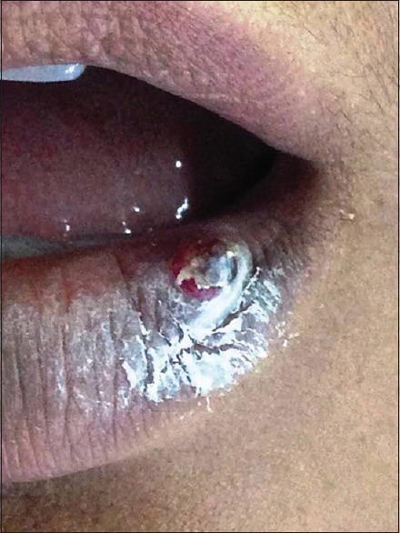
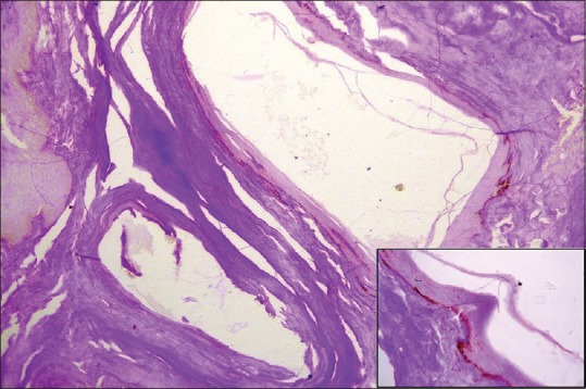
Keratoacanthoma (KA) is a benign epithelial tumor which presents clinically as a proliferating dome-shaped keratin-filled crater. The lesion occurs predominantly upon sun-exposed areas of the body and is known to arise from hair follicle. Actinic rays are a major contributing factor in the etiology. It simulates grossly as well as microscopically a low-grade squamous cell carcinoma. KAs occur habitually on the mucous membrane as well, but their origin in these cases is debatable, owing to the lack of hair follicles in these sites. Our report is an attempt to demonstrate the cells that could be responsible for initiation of this lesion on the oral mucosa.

Keywords: Ectopic sebaceous gland; follicular infundibulum; keratoacanthoma.


Conflict of interest statement

There are no conflicts of interest.

Figures

Figure 1
Figure 1
Clinical image shows noduloulcerative growth with keratin plugging seen on left vermilion of the lower lip
Figure 2
Figure 2
Histopathological image shows hyperkeratotic epithelium with parakeratin plugging overlying connective tissue stroma with epithelial proliferations superficially in the stroma (H&E, ×40)
Figure 3
Figure 3
Histopathological image shows epithelial tissue entrapment with cystic degeneration in the center and keratinization (H&E, ×100). Inset (H&E, ×400)
Figure 4
Figure 4
Histopathological image shows periodic acid–Schiff-stained section showing negative staining for glycogen granules (H&E, ×100). Inset (PAS, ×400)

References

    1. Karnauchow PN. A lesion simulating squamous carcinoma of the lip (keratoacanthoma) Can Med Assoc J. 1958;78:346–8. - PMC - PubMed
    1. Fisher BK, Elliott GB. On the origin of keratoacanthoma: Reflections on an unusual lesion. Can Med Assoc J. 1965;93:272–3. - PMC - PubMed
    1. Ramos LM, Cardoso SV, Loyola AM, Rocha MA, Durighetto-Júnior AF. Keratoacanthoma of the inferior lip: Review and report of case with spontaneous regression. J Appl Oral Sci. 2009;17:262–5. - PMC - PubMed
    1. Chauhan A, Chaudhary S, Agnihotri PG, Aadithya B. A solitary crateriform ulcer of the lower lip: A case report with review of literature. Indian J Dermatol. 2011;56:435–8. - PMC - PubMed
    1. Patil PB, Rathor V, Venkatraman S, Saxena S, Kamarthi N. Solitary keratoacanthoma involving upper lip: A diagnostic dilemma- case report and a brief review. J Clin Exp Dent. 2010;2:e34–7.

Publication types