

The potential of enriched mesenchymal stem cells with neural crest cell phenotypes as a cell source for regenerative dentistry
- PMID: 28479933
- PMCID: PMC5405184
- DOI: 10.1016/j.jdsr.2016.09.001
The potential of enriched mesenchymal stem cells with neural crest cell phenotypes as a cell source for regenerative dentistry
Abstract
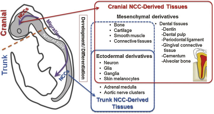
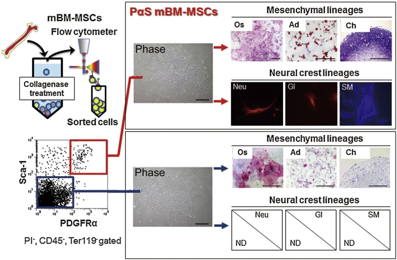
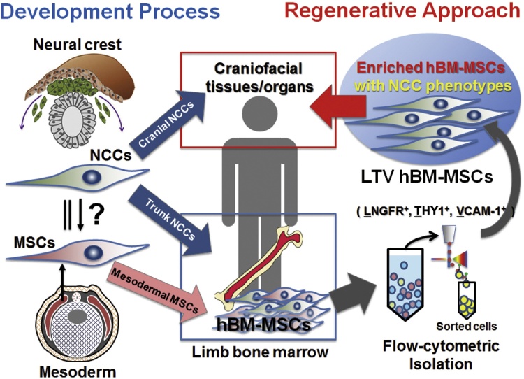
Effective regenerative treatments for periodontal tissue defects have recently been demonstrated using mesenchymal stromal/stem cells (MSCs). Furthermore, current bioengineering techniques have enabled de novo fabrication of tooth-perio dental units in mice. These cutting-edge technologies are expected to address unmet needs within regenerative dentistry. However, to achieve efficient and stable treatment outcomes, preparation of an appropriate stem cell source is essential. Many researchers are investigating the use of adult stem cells for regenerative dentistry; bone marrow-derived MSCs (BM-MSCs) are particularly promising and presently used clinically. However, current BM-MSC isolation techniques result in a heterogeneous, non-reproducible cell population because of a lack of identified distinct BM-MSC surface markers. Recently, specific subsets of cell surface markers for BM-MSCs have been reported in mice (PDGFRα+ and Sca-1+) and humans (LNGFR+, THY-1+ and VCAM-1+), facilitating the isolation of unique enriched BM-MSCs (so-called "purified MSCs"). Notably, the enriched BM-MSC population contains neural crest-derived cells, which can differentiate into cells of neural crest- and mesenchymal lineages. In this review, characteristics of the enriched BM-MSCs are outlined with a focus on their potential application within future regenerative dentistry.
Keywords: Bone marrow-derived mesenchymal stem cell; Enriched/purified mesenchymal stem cell; Flow cytometric isolation; Neural crest cell; Regenerative dentistry.
Figures
Similar articles
-
LNGFR+THY-1+ human pluripotent stem cell-derived neural crest-like cells have the potential to develop into mesenchymal stem cells.Differentiation. 2016 Dec;92(5):270-280. doi: 10.1016/j.diff.2016.04.003. Epub 2016 May 10. Differentiation. 2016. PMID: 27178356
-
Comparative Craniofacial Bone Regeneration Capacities of Mesenchymal Stem Cells Derived from Human Neural Crest Stem Cells and Bone Marrow.ACS Biomater Sci Eng. 2021 Jan 11;7(1):207-221. doi: 10.1021/acsbiomaterials.0c00878. Epub 2020 Dec 9. ACS Biomater Sci Eng. 2021. PMID: 33455206
-
Different Sources of Mesenchymal Stem Cells for Tissue Regeneration: A Guide to Identifying the Most Favorable One in Orthopedics and Dentistry Applications.Int J Mol Sci. 2022 Jun 6;23(11):6356. doi: 10.3390/ijms23116356. Int J Mol Sci. 2022. PMID: 35683035 Free PMC article. Review.
-
Induced Pluripotent Stem Cell-Derived Mesenchymal Stromal Cells Are Functionally and Genetically Different From Bone Marrow-Derived Mesenchymal Stromal Cells.Stem Cells. 2019 Jun;37(6):754-765. doi: 10.1002/stem.2993. Epub 2019 Mar 6. Stem Cells. 2019. PMID: 30779868 Free PMC article.
-
Isolation and characterization of primary bone marrow mesenchymal stromal cells.Ann N Y Acad Sci. 2016 Apr;1370(1):109-18. doi: 10.1111/nyas.13102. Ann N Y Acad Sci. 2016. PMID: 27270495 Review.
Cited by
-
Novel Mesenchymal Stem Cell Spheroids with Enhanced Stem Cell Characteristics and Bone Regeneration Ability.Stem Cells Transl Med. 2022 Apr 29;11(4):434-449. doi: 10.1093/stcltm/szab030. Stem Cells Transl Med. 2022. PMID: 35267026 Free PMC article.
-
miR-148b-3p, miR-337-5p and miR-423-5p expression in alveolar ridge atrophy and their roles in the proliferation and apoptosis of OMMSCs.Exp Ther Med. 2018 Dec;16(6):5334-5342. doi: 10.3892/etm.2018.6850. Epub 2018 Oct 11. Exp Ther Med. 2018. PMID: 30542492 Free PMC article.
-
A Shaking-Culture Method for Generating Bone Marrow Derived Mesenchymal Stromal/Stem Cell-Spheroids With Enhanced Multipotency in vitro.Front Bioeng Biotechnol. 2020 Oct 20;8:590332. doi: 10.3389/fbioe.2020.590332. eCollection 2020. Front Bioeng Biotechnol. 2020. PMID: 33195156 Free PMC article.
-
Effect of Long-Term 3D Spheroid Culture on WJ-MSC.Cells. 2021 Mar 24;10(4):719. doi: 10.3390/cells10040719. Cells. 2021. PMID: 33804895 Free PMC article.
-
Investigate the Odontogenic Differentiation and Dentin-Pulp Tissue Regeneration Potential of Neural Crest Cells.Front Bioeng Biotechnol. 2020 Jun 5;8:475. doi: 10.3389/fbioe.2020.00475. eCollection 2020. Front Bioeng Biotechnol. 2020. PMID: 32582651 Free PMC article.
References
-
- Egusa H., Sonoyama W., Nishimura M., Atsuta I., Akiyama K. Stem cells in dentistry – Part I: stem cell sources. J Prosthodont Res. 2012;56:151–165. - PubMed
-
- Masaki C., Nakamoto T., Mukaibo T., Kondo Y., Hosokawa R. Strategies for alveolar ridge reconstruction and preservation for implant therapy. J Prosthodont Res. 2015;59:220–228. - PubMed
-
- Kaku M., Akiba Y., Akiyama K., Akita D., Nishimura M. Cell-based bone regeneration for alveolar ridge augmentation – cell source, endogenous cell recruitment and immunomodulatory function. J Prosthodont Res. 2015;59:96–112. - PubMed
-
- Li P., Honda Y., Arima Y., Yasui K., Inami K., Nishiura A. Interferon-γ enhances the efficacy of autogenous bone grafts by inhibiting postoperative bone resorption in rat calvarial defects. J Prosthodont Res. 2016;60:167–176. - PubMed
-
- Verhoeven J.W., Ruijter J., Cune M.S., Terlou M., Zoon M. Onlay grafts in combination with endosseous implants in severe mandibular atrophy: one year results of a prospective, quantitative radiological study. Clin Oral Implants Res. 2000;11:583–594. - PubMed
Publication types
LinkOut - more resources
Full Text Sources
Other Literature Sources
Research Materials
Miscellaneous
