

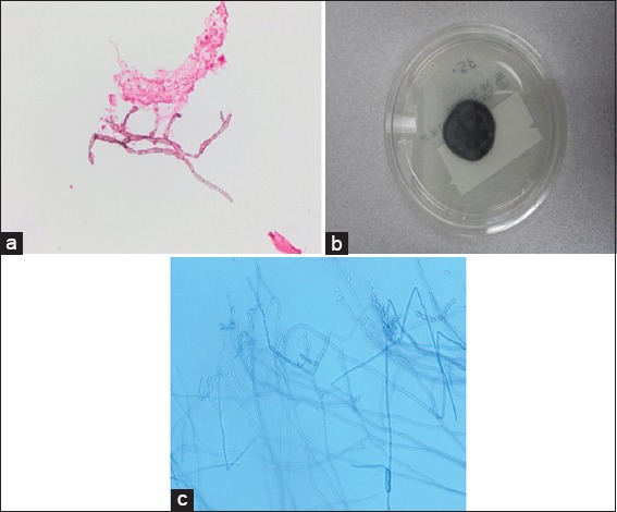
Fungal brain abscess caused by "Black Mold" (Cladophialophora bantiana) - A case report of successful treatment with an emphasis on how fungal brain abscess may be different from bacterial brain abscess
- PMID: 28480108
- PMCID: PMC5402337
- DOI: 10.4103/sni.sni_448_16
Fungal brain abscess caused by "Black Mold" (Cladophialophora bantiana) - A case report of successful treatment with an emphasis on how fungal brain abscess may be different from bacterial brain abscess
Abstract
Background: Central nervous system infection with Cladophialophora bantiana (Black Mold) is rare. It carries a high mortality rate, that is more than 70%, despite multimodal therapy.
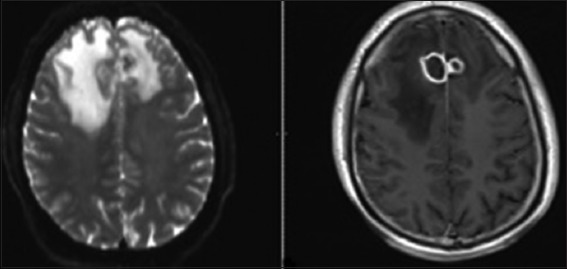
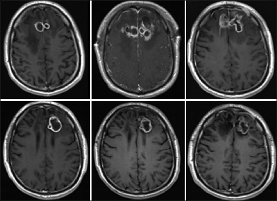
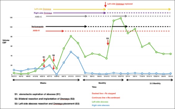
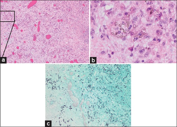
Case description: We present a rare case of "black mold" fungal brain abscess that was successfully treated with good patient outcome. The case is unusual because there were two fungal brain abscesses located bilaterally symmetrically in the mesial frontal lobes, and the response to different treatment strategies was well documented by over 25 magnetic resonance imaging (MRI) scans. Initial attempts to treat these lesions by repeated surgical excision and systemic amphotericin B was followed by continued growth rather than resolution. We realized that the application of treatment principles learned from bacterial brain abscess may not transpose intuitively to the treatment of fungal brain abscess. Therefore, a new treatment strategy was adopted that avoided further attempts at resection in favor of long-term oral voriconazole and repeated intracavitary aspiration and instillation of amphotericin B on an outpatient basis. Without further resection, the lesions stabilized and the aspirates eventually sterilized, however, the enhancing capsule never resolved on MRI scans. All treatment was stopped after 1 year. The apparently sterilized lesions have been followed for an additional 3 years without further growth, and the patient remains functionally, intellectually, and behaviorally normal.
Conclusion: We conclude that, in the case of fungal abscess, it may be preferable to sterilize the lesion in situ rather than attempting to achieve resolution on imaging studies by repeated surgical resection of the capsule which can be counterproductive. This strategy accepts that the capsule may be important to the patient's immune defense against the fungus. Helping that defense barrier with intracapsular and systemic antifungal agents, rather than capsular removal, may be the better strategy for patients in whom early aggressive resection has failed. The basis for the apparent differences in the response of fungal versus bacterial brain abscess to surgical resection is discussed in the light of pathological findings from this and other cases.
Keywords: Abscess; antifungal; black mold; brain; excision.
Conflict of interest statement
There are no conflicts of interest.
Figures
References
-
- Adler DE, Milhorat TH, Miller JI. Treatment of rhinocerebral mucormycosis with intravenous interstitial, and cerebrospinal fluid administration of amphotericin B: Case report. Neurosurgery. 1998;42:644–8. - PubMed
-
- Al-Abdely HM, Alkhunaizi AM, Al-Tawfiq JA, Hassounah M, Rinaldi MG, Sutton DA. Successful therapy of cerebral phaeohyphomycosis due to Ramichloridium mackenziei with the new triazole posaconazole. Med Mycol. 2005;43:91–5. - PubMed
-
- Banerjee U, Mohapatra AK, Sarkar C, Chaudhery R. Cladosporiosis (cerebral phaeohyphomycosis) of brain--a case report. Mycopathologia. 1989;105:163–6. - PubMed
-
- Biggs PJ, Allen RL, Powers JM, Holley HP., Jr Phaeohyphomycosis complicating compound skull fracture. Surg Neurol. 1986;25:393–6. - PubMed
Publication types
LinkOut - more resources
Full Text Sources
Other Literature Sources