Review
. 2017 Apr 5:7:15.
doi: 10.4103/jcis.JCIS_6_17. eCollection 2017.

Large Bowel Obstruction in the Emergency Department: Imaging Spectrum of Common and Uncommon Causes

Affiliations
Review

Large Bowel Obstruction in the Emergency Department: Imaging Spectrum of Common and Uncommon Causes

Subramaniyan Ramanathan et al. J Clin Imaging Sci. .

Abstract

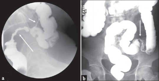
Although large bowel obstruction (LBO) is less common than small bowel obstruction, it is associated with high morbidity and mortality due to delayed diagnosis and/or treatment. Plain radiographs are sufficient to diagnose LBO in a majority of patients. However, further evaluation with multidetector computed tomography (MDCT) has become the standard of care to identify the site, severity, and etiology of obstruction. In this comprehensive review, we illustrate the various causes of LBO emphasizing the role of MDCT in the initial diagnosis and detection of complications along with the tips to differentiate from disease which can mimic LBO.

Keywords: Diverticulitis; large bowel obstruction; multidetector computed tomography; neoplastic; pseudo-obstruction; volvulus.


Conflict of interest statement

There are no conflicts of interest.

Figures

Figure 1
Figure 1
Colorectal cancer: (a and b) Axial contrast-enhanced computed tomography images in a 60-year-old male patient with colorectal cancer presenting with weight loss and chronic abdominal pain demonstrate marked distension of transverse colon and splenic flexure (long arrow in a) with associated short segment mural thickening of proximal descending colon with abrupt transition (short arrow) to a normal appearing descending colon (long arrow in b). The large bowel dilatation and abrupt transition are better depicted on the coronal image (c).
Figure 2
Figure 2
Extra colonic malignancies: (a and b) Axial contrast-enhanced computed tomography images in a 56-year-old female patient with history of ovarian cancer presenting with abdominal distension demonstrate ascites, peritoneal thickening and omental caking consistent with peritoneal carcinomatosis (arrow in a). There is marked distension of the proximal large bowel and fecal loading of the descending colon. The pelvic peritoneal metastatic deposits caused extrinsic narrowing of the sigmoid colon (arrow in b) and accounted for the large bowel obstruction. The large bowel dilatation and transition point in the sigmoid colon are better depicted on the coronal image (c).
Figure 3
Figure 3
Extracolonic malignancy: Axial (a and b) and coronal (c) contrast-enhanced computed tomography images in a 73-year-old man presenting with weight loss and worsening left upper quadrant abdominal pain demonstrate a heterogeneous mass arising from the pancreatic tail which is contiguously infiltrating the splenic flexure of colon (arrow in a). There is marked distension of ascending and transverse colon and splenic flexure (asterisk) with decompressed descending colon (arrows in b and c). The large bowel obstruction resulted from contiguous infiltration of the splenic flexure from pancreatic tail cancer.
Figure 4
Figure 4
Large bowel obstruction secondary to diverticular stricture. Plain radiographs of the abdomen, supine and erect views (a and b) in a 45-year-old man presenting to the emergency department with severe abdominal pain and lack of bowel movements demonstrate markedly dilated loops of large bowel compatible with large bowel obstruction. Axial (c) and coronal (d) contrast-enhanced computed tomography images demonstrate dilated large bowel loops (arrow in c) with associated concentric thickening and stricture (arrow in d) of the distal descending colon without discrete mass. Single column barium enema (e) performed subsequently better demonstrates the stricture (short arrow) and an additional fistula (long arrow). Axial computed tomography image (f) performed an year earlier demonstrates changes of acute diverticulitis which eventually led to stricture and large bowel obstruction.
Figure 5
Figure 5
Sigmoid volvulus: (a) Erect plain abdominal radiograph in a 58-year-old man presenting with abdominal distension and severe lower abdominal pain demonstrates marked distension of the sigmoid colon with a typical coffee-bean sign (arrows) which is highly characteristic of sigmoid volvulus. A rubber tube has been placed for decompression. Axial contrast-enhanced computed tomography images (b and c) in another patient with similar presentation demonstrates the whirl pattern (arrow in b) and the characteristic bird beak appearance (arrow in c). The redundant and dilated sigmoid colon (asterisk) is better depicted on the coronal image (d) with characteristic beak appearance (arrow).
Figure 6
Figure 6
Cecal volvulus: (a) Erect plain radiograph of the abdomen in a 65-year-old male patient presenting with severe abdominal pain demonstrates mildly dilated loops of small bowel (short arrows) in the right hemi abdomen and a markedly dilated gas filled structure in the left upper quadrant (long arrow) which is compressing the stomach which contains nasogastric tube. Axial contrast enhanced computed tomography images (b and c) demonstrate a large air and fluid filled structure in the left abdomen (asterisk) with absence of cecum in its normal location. A small amount of fluid (arrow) is noted secondary to early ischemia. The enteric contrast is noted to opacify only the small bowel. Imaging findings are concerning for cecal volvulus which was subsequently confirmed at surgery.
Figure 7
Figure 7
Colonic lipoma complicated by intussusception. Colonic lipoma complicated by intussusception. Axial (a) contrast-enhanced computed tomography in a 40-year-old man demonstrates small colonic lipoma (arrow). Follow-up computed tomography 5 years later when the patient presented clinically with symptoms of abdominal pain and subacute intestinal obstruction. Axial (b-d) contrast-enhanced computed tomography images demonstrate interval significant growth of the lipoma (short arrows) with associated colonic intussusception (long arrows).
Figure 8
Figure 8
Enterolith: (a and b) Erect and supine plain abdominal radiograph in a 56-year-old man presenting with abdominal distension, vomiting, and severe lower abdominal pain demonstrates dilatation of the small and large bowel loops with associated air-fluid levels concerning for obstruction. A lamellated calcific shadow (arrow) is noted in the left lower quadrant. Computed tomography scan (c-e) performed subsequently confirms an enterolith (arrows) in the distal descending colon which resulted in bowel obstruction. No mass was identified and the patient was managed conservatively with endoscopic fragmentation and removal of the enterolith. Plain abdominal radiograph performed subsequently demonstrated complete removal of the enterolith and interval improvement of bowel obstruction (f).
Figure 9
Figure 9
Fecal impaction: (a and b) Erect and supine plain abdominal radiograph in a 75-year-old man presenting with abdominal distension and worsening lower abdominal pain demonstrates dilatation of the large bowel loops. Computed tomography scan (c and d) performed subsequently demonstrates large amount of stool in the sigmoid colon and rectum (arrows) which resulted in large bowel dilatation. No mass was identified and the patient was managed conservatively with enemas. Repeat computed tomography performed subsequently demonstrates resolution of the impacted feces with residual fluid in the sigmoid colon (e).
Figure 10
Figure 10
Ogilvie's syndrome: (a) Plain abdominal radiograph in a 61-year-old man presenting with abdominal distension demonstrates marked distension of the colon (arrow). Computed tomography scan (b-d) performed subsequently reveal distended colon (arrows). There is no evidence of small bowel dilatation or obstructing lesion. The patient was managed conservatively.
Figure 11
Figure 11
Adynamic ileus in a 65-year-old man with diabetes presenting with diffuse intermittent abdominal pain and found to have sever hypokalemia. (a) Plain abdominal radiograph shows diffuse dilatation of colon. Axial (b-d) contrast-enhanced computed tomography shows diffuse dilatation of both small bowel (long arrows) and colon (short arrows) all the way to rectum without transition.
Figure 12
Figure 12
Toxic megacolon in a 55 year old man with known ulcerative colitis. (a) Plain abdominal radiograph shows dilatation of transverse colon and splenic flexure with loss of haustrations. Coronal (b) CT images show the dilatation of splenic flexure with loss of haustrations (arrows). Axial (c and d) CT images demonstrate the changes of acute colitis in the cecum (arrow in c) and rectosigmoid (arrow in d).
Figure 13
Figure 13
Stricture: (a) Single column barium enema in a 59-year-old man who underwent pelvic irradiation demonstrates a long segment irregular stricture of the rectosigmoid (long arrow). Also noted is a colocolic fistula (short arrow) which is another potential complication of radiation therapy. Barium enema in another patient with history of ischemic colitis demonstrates a short segment smooth stricture (arrow) at the junction of the sigmoid and descending colon (b).

References

    1. Taourel P, Kessler N, Lesnik A, Pujol J, Morcos L, Bruel JM. Helical CT of large bowel obstruction. Abdom Imaging. 2003;28:267–75. - PubMed
    1. Yeo HL, Lee SW. Colorectal emergencies: Review and controversies in the management of large bowel obstruction. J Gastrointest Surg. 2013;17:2007–12. - PubMed
    1. Gore RM, Levine MS, editors. Textbook of Gastrointestinal Radiology. 3rd ed. Philadelphia, PA: Saunders, Elsevier; 2008.
    1. Horton KM, Corl FM, Fishman EK. CT evaluation of the colon: Inflammatory disease. Radiographics. 2000;20:399–418. - PubMed
    1. Frager D, Rovno HD, Baer JW, Bashist B, Friedman M. Prospective evaluation of colonic obstruction with computed tomography. Abdom Imaging. 1998;23:141–6. - PubMed