

Theoretical diagnosis of emphysema by aerosol bolus inhalation
- PMID: 28480190
- PMCID: PMC5401684
- DOI: 10.21037/atm.2017.03.28
Theoretical diagnosis of emphysema by aerosol bolus inhalation
Abstract
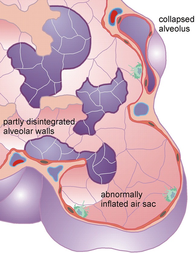
Background: The present contribution deals with the theoretical description of aerosol bolus dispersion in lungs being affected by different manifestations of emphysema. The work constructs the hypothesis that each manifestation of emphysema exhibits specific properties with regard to the dispersion of inhaled and exhaled aerosol boluses as well as the deposition of particles from the aerosol pulse.
Methods: For an appropriate simulation of single emphysematous manifestations, a previously developed model assuming (I) a random variation of alveolar diameters, (II) an exact localization of diseased structures, and (III) a realistic balance between alveolar air volume and number of air sacs was applied. Dispersion of inhaled and exhaled aerosol boluses was simulated by using the mathematical concept of effective diffusivities. Computations were conducted for an average adult lung (FRC =3,300 mL), symmetric breath-cycles with a length 8 s, and inhalation flow rates of 250 mL/s. Particles used for the model predictions had a uniform diameter of 0.84 µm and a density of 1 g/cm3.
Results: According to the theoretical data obtained from the model highest aerosol bolus dispersion may be observed in lungs affected by panacinar and bullous emphysema, whereas centriacinar and paraseptal emphysema cause a significant reduction of the phenomenon. Also other statistical parameters exhibit partly remarkable differences among the studied manifestations. Particle deposition in lungs affected by bullous emphysema falls below that of lungs impaired by the other types of emphysema by 2%-50%.
Conclusions: From the hypothetical results presented in this study it may be concluded that aerosol bolus inhalation bears a certain potential for the diagnosis of emphysematous structures and, if applied with sufficient accuracy, also for the distinction of single manifestations of emphysema. For a successful use of the technique, however, all statistical bolus parameters and particle deposition have to be subjected to a detailed evaluation.
Keywords: Aerosol bolus; dispersion; emphysema; lung; stochastic model.
Conflict of interest statement
Conflicts of Interest: The author has no conflicts of interest to declare.
Figures


References
-
- Fitzpatrick M. Studies on human pulmonary connective tissue. Am Rev Respir Dis 1967;96:254-65. - PubMed
LinkOut - more resources
Full Text Sources
Other Literature Sources