. 2017 May;22(3):e12447.
doi: 10.1111/anec.12447.

2017 ISHNE-HRS expert consensus statement on ambulatory ECG and external cardiac monitoring/telemetry

Affiliations

2017 ISHNE-HRS expert consensus statement on ambulatory ECG and external cardiac monitoring/telemetry

Jonathan S Steinberg et al. Ann Noninvasive Electrocardiol. 2017 May.

Abstract

Ambulatory ECG (AECG) is very commonly employed in a variety of clinical contexts to detect cardiac arrhythmias and/or arrhythmia patterns which are not readily obtained from the standard ECG. Accurate and timely characterization of arrhythmias is crucial to direct therapies that can have an important impact on diagnosis, prognosis or patient symptom status. The rhythm information derived from the large variety of AECG recording systems can often lead to appropriate and patient-specific medical and interventional management. The details in this document provide background and framework from which to apply AECG techniques in clinical practice, as well as clinical research.

Keywords: Holter; ambulatory ECG monitoring; event monitor; loop recorder; telemetry; transtelephonic.


Figures

Figure 1
Figure 1
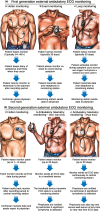
Types of AECG monitors currently available in clinical practice. (a) Holter, event, and loop monitoring; (b) patch‐type extended Holter and ambulatory telemetry monitoring AECG, ambulatory external electrocardiographic; ECG, electrocardiographic. Figure illustration by Craig Skaggs. Reproduced with permission from Mittal et al. (2011).
Figure 2
Figure 2
Holter lead configurations. (a) Mason‐Likar 12‐lead system for continuous ECG monitoring. Electrode placements are as follows: RA: 2nd intercostal space right side midclavicular, LA 2nd intercostal space left side midclavicular, LL below 8th intercostal left side midclavicular line, RL: upper sternum, V1: 4th right intercostal space at the sternal border, V2: 4th left intercostal space at the sternal border, V3: between V2 and V4, V4: 5th left intercostal space at the midclavicular line, V5: 5th left intercostal space at the anterior axillary line, V6: 5th left intercostal space at the midaxillary line; (b) EASI reduced leads system. EASI is a reduced lead system suitable for continuous ECG monitoring. It is an alternative to both the commonly used 5‐electrode monitoring system and the traditional 10‐electrode Mason‐Likar 12‐lead ECG system. The EASI lead configuration enables continuous reconstructed 12‐lead ECG ambulatory monitoring using only five electrodes. The EASI 12‐lead ECG is derived from a set of four recording electrodes and one reference electrode. The placement of these leads is as follows: E: lower extreme of the sternum, A: left mid‐axillary line, same transverse line as E, S: sternal manubrium, I: right mid‐axillary line, same transverse line as E, R: fifth electrode is the body potential reference and can be placed anywhere on the torso
Figure 3
Figure 3
Examples of ECG artifacts (a) The bottom channel shows recording artifact that may simulate atrial flutter/fibrillation. However, careful analysis of the upper channel shows sinus rhythm with clear P waves. The presence of an irregular rhythm secondary to both sinus arrhythmia and occasional premature atrial beats adds to difficulty in making the correct diagnosis from the recording in the bottom channel alone. Figures (b and c) are two distinct examples of artifacts from the same Holter recording that may simulate ventricular tachyarrhythmia. In both tracings, the artifacts are more prominent in one of the two illustrated channels but not the other, making correct interpretation feasible. Normal QRS complexes are marked by arrows at the channel with prominent artifacts. Reproduced with permission from El‐Sherif & Turitto (2015)
Figure 4
Figure 4
Electronic Health Record (EHR) integration workflow. The EHR contains information about the patient, which is captured at registration. In this example, the practice is using a third‐party provider for ambulatory electrocardiographic monitoring. The EHRPipe is an interface of interrogation engine that serves as a bidirectional bridge between this provider and the EHR system (courtesy of ScottCare, Inc.)

References

    1. Ackermans, P. A. , Solosko, T. A. , Spencer, E. C. , Gehman, S. E. , Nammi, K. , Engel, J. , & Russell, J. K. (2012). A user‐friendly integrated monitor‐adhesive patch for long‐term ambulatory electrocardiogram monitoring. Journal of Electrocardiology, 45, 148–153. - PubMed
    1. Adabag, A. S. , Casey, S. A. , Kuskowski, M. A. , Zenovich, A. G. , & Maron, B. J. (2005). Spectrum and prognostic significance of arrhythmias on ambulatory Holter electrocardiogram in hypertrophic cardiomyopathy. Journal of the American College of Cardiology, 45, 697–704. - PubMed
    1. Adabag, A. S. , Maron, B. J. , Appelbaum, E. , Harrigan, C. J. , Buros, J. L. , Gibson, C. M. , … Maron, M. S. (2008). Occurrence and frequency of arrhythmias in hypertrophic cardiomyopathy in relation to delayed enhancement on cardiovascular magnetic resonance. Journal of the American College of Cardiology, 51, 1369–1374. - PubMed
    1. Ajami, S. , & Teimouri, F. (2015). Features and application of wearable biosensors in medical care. Journal of Research in Medical Sciences, 20, 1208–1215. - PMC - PubMed
    1. Alboni, P. , Botto, G. , Baldi, N. , Luzi, M. , Russo, V. , Gianfranchi, L. , … Gaggioli, G. (2004). Outpatient treatment of recent‐onset atrial fibrillation with the “Pill‐in‐the‐Pocket” approach. New England Journal of Medicine, 351, 2383–2391. - PubMed

Publication types