Review
. 2017 May;19(2):166-187.
doi: 10.5853/jos.2016.01368. Epub 2017 May 8.

Emerging Roles of microRNAs in Ischemic Stroke: As Possible Therapeutic Agents

Affiliations
Review

Emerging Roles of microRNAs in Ischemic Stroke: As Possible Therapeutic Agents

Seyed Esmaeil Khoshnam et al. J Stroke. 2017 May.

Abstract

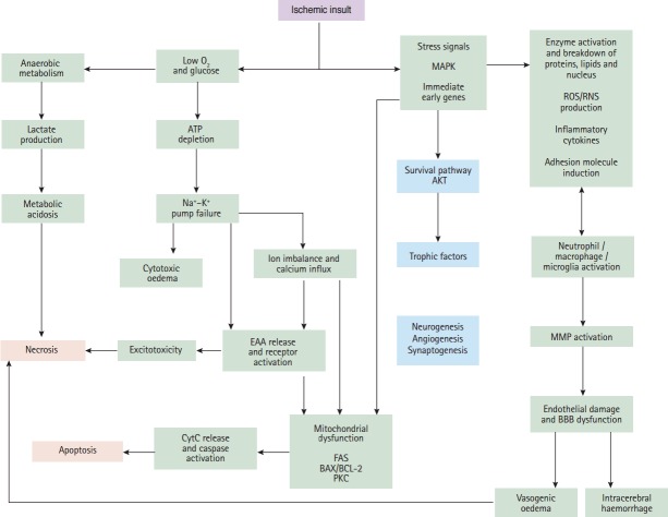
Stroke is one of the leading causes of death and physical disability worldwide. The consequences of stroke injuries are profound and persistent, causing in considerable burden to both the individual patient and society. Current treatments for ischemic stroke injuries have proved inadequate, partly owing to an incomplete understanding of the cellular and molecular changes that occur following ischemic stroke. MicroRNAs (miRNA) are endogenously expressed RNA molecules that function to inhibit mRNA translation and have key roles in the pathophysiological processes contributing to ischemic stroke injuries. Potential therapeutic areas to compensate these pathogenic processes include promoting angiogenesis, neurogenesis and neuroprotection. Several miRNAs, and their target genes, are recognized to be involved in these recoveries and repair mechanisms. The capacity of miRNAs to simultaneously regulate several target genes underlies their unique importance in ischemic stroke therapeutics. In this Review, we focus on the role of miRNAs as potential diagnostic and prognostic biomarkers, as well as promising therapeutic agents in cerebral ischemic stroke.

Keywords: Ischemia; MicroRNAs; Stroke.


Conflict of interest statement

The authors have no financial conflicts of interest.

Figures

Figure 1.
Figure 1.
Critical events in the ischemic cascade. Following ischaemia, the deprivation of oxygen and glucose to the brain lead to loss of ATP (energy loss) and ion pump failure. The loss of ion concentration gradients causes cytotoxic oedema and releasing of excitatory amino acids (EAAs). Following reduced glucose availability cell aerobic metabolism switches to anaerobic, resulting in metabolic acidosis. All of these events lead to cell death, or necrosis. Ischaemia also causes the upregulation and activation of many immediate early genes and stress signals, which lead to inflammatory responses, cell apoptosis and, subsequently, activation of matrix metalloproteinases (MMPs) as a damaging protease which can lead to the brain oedema and haemorrhage. Following ischaemia, AKT kinase activation and upregulation of trophic factors set the stage for recovery and repair mechanisms which including neurogenesis, synaptogenesis and angiogenesis. AKT, protein kinase B; MAPK, mitogen-activated protein kinase; ROS/RNS, reactive oxygen species/reactive nitrogen species; ATP, adenosine triphosphate; EAA, excitatory amino acids; CytC, cytochrome c; FAS, the cell-surface Fas receptor; PKC, protein kinase C; BBB, blood brain barrier.
Figure 2.
Figure 2.
MicroRNAs involved in detrimental (purple boxes) and protective pathways (blue boxes) are activated by ischemic stroke. Cerebral ischemia, while activating detrimental pathways, also triggers some organized responses that counteract tissue injury. Post-ischemic oxidative stress triggers an oxidant and antioxidant responses via different factors which are inhibited by microRNAs. Oxidative agents that are inhibited by microRNAs, including reactive oxygen/nitrogen species (ROS/RNS), cyclooxygenase 2 (COX2), hydrogen peroxide (H2O2), malondialdehyde (MDA) and methane dicarboxylic aldehyde (MEDA). The antioxidant response which is inhibited by microRNAs containing transcription factor Nrf2 and superoxide dismutase (SOD). Following ischemia, inflammation is increased by production of matrix metalloproteinases (MMP-9) to infiltrate the BBB, and activation of pro-inflammatory genes such as interleukin-1 (IL-1α and IL-1β), IL-6, tumor necrosis factor α (TNF-α) and nuclear factor-κB, (NF-κB), as well as an activation of innate immune responses (microglia cells) and toll-like receptors (TLR4). Inflammation is mitigated by production of anti-inflammatory cytokines like such as IL-10. microRNAs could affect post-ischemic inflammatory and anti-inflammatory factors. Excitotoxicity associated with glutamate receptor activation can be counterbalance via glutamate transporter (GLT1) and NMDA (containing subunit NR2A), while glutamate receptors GluR2 and NMDA (containing subunit NR2B) exacerbate excitotoxic injuries. microRNAs inhibit those factors that contribute in the excitotoxicity. The detrimental effects of post-ischemic apoptosis are antagonized by activation and expression of antiapoptotic factors such as; Bcl-2, Bcl2L11, Bcl-w, Mcl-1 and the heat shock proteins family (HSPA12B). Hence, deleterious effects of apoptosis are induced by expression of caspase 3, activation of cell surface death receptors (Fas) and its ligand (FasL), and activation of p53, inhibitory member of the apoptosis-stimulating proteins of the p53 family (iASPP). There are some microRNAs which modulate the detrimental effects of post-ischemic apoptosis. SOCS1, suppressor of cytokine signaling 1; MyD88, myeloid differentiation primary response gene 88; iNOS, inducible nitric oxide synthase; Nrf2, nuclear factor erythroid-2 related factor 2; PUMA, p53 upregulated modulator of apoptosis; GLT-1, glutamate transporter-1; GluR2, glutamate receptor-2; FAP-1, Fas associated protein-tyrosine phosphatase 1.
Figure 3.
Figure 3.
Overview of processes involved in ischemic stroke and high potential therapeutic microRNAs. Cerebral ischemia includes several injurious mechanisms (excitotoxicity, oxidative stress, inflammation and apoptosis) to confer neuronal injury. Potential therapeutic areas to compensate for these pathogenic process include promoting angiogenesis, neurogenesis and neuroprotective recovery and repair mechanisms.

Similar articles

Cited by

References

    1. Heron M. Deaths: leading causes for 2004. Natl Vital Stat Rep. 2007;56:1–96. - PubMed
    1. Centers for Disease Control and Prevention (CDC) Prevalence of disabilities and associated health conditions among adults--United Sstates, 1999. Morb Mortal Wkly Rep. 2001;50:120–125. - PubMed
    1. World Health Organization . The World health report 2004: changing history. Geneva: World Health Organization; 2004.
    1. Mukherjee D, Patil CG. Epidemiology and the global burden of stroke. World Neurosurg. 2011;76(6 Suppl):S85–S90. - PubMed
    1. Beal CC. Gender and stroke symptoms: a review of the current literature. J Neurosci Nurs. 2010;42:80–87. - PubMed