

Disruption of Estrogen Receptor Alpha in Rats Results in Faster Initiation of Compensatory Regeneration Despite Higher Liver Injury After Carbon Tetrachloride Treatment
- PMID: 28481132
- PMCID: PMC5535772
- DOI: 10.1177/1091581817706067
Disruption of Estrogen Receptor Alpha in Rats Results in Faster Initiation of Compensatory Regeneration Despite Higher Liver Injury After Carbon Tetrachloride Treatment
Abstract
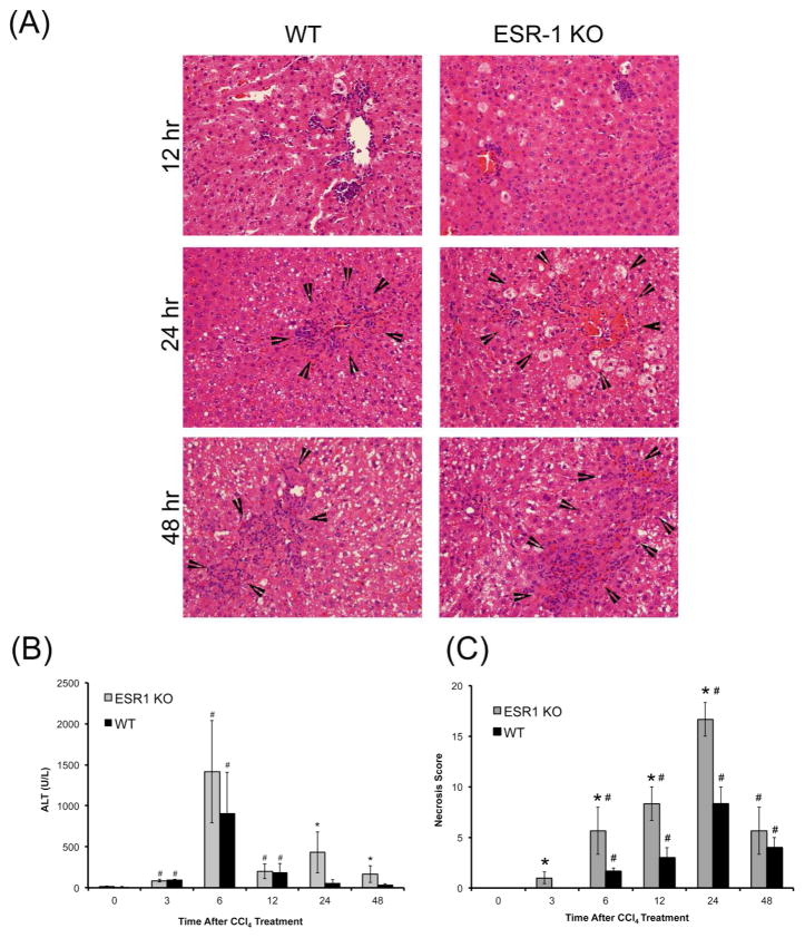
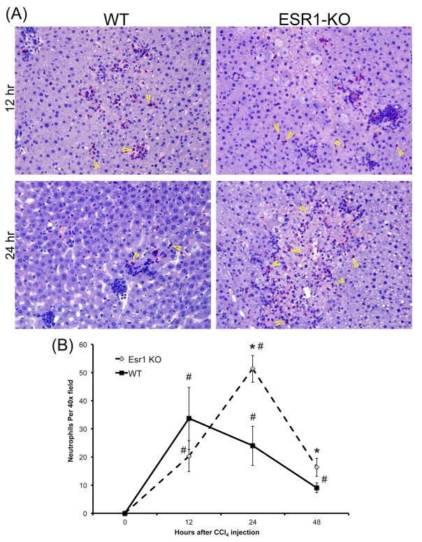
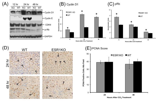
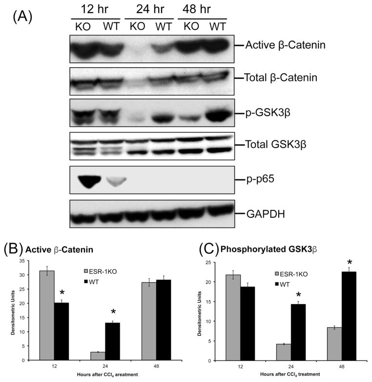
Estrogen receptor alpha (ESR1) is 1 of the 2 intracellular receptors for estrogen and is expressed by hepatocytes in the liver. The role of ESR1 in the regulation of toxicant-induced liver injury and compensatory regeneration is not completely clear. We investigated the role of ESR1 in liver regeneration after carbon tetrachloride (CCl4)-induced liver injury using wild type (WT) and ESR1 knockout (ESR1-KO) rats. Adult female WT and ESR1-KO rats were treated with 1 mL/kg CCl4 and euthanized over a time course of 0 to 48 hours. Liver injury measured by serum alanine amino transaminase, and histopathological analysis showed significantly higher liver injury in ESR1-KO as compared to WT rats. Hematoxylin and eosin staining revealed 2-fold higher necrosis and significant inflammatory cell infiltration in ESR1-KO rats. Chloracetate esterase staining revealed higher neutrophil infiltration in ESR1-KO rat livers. Interestingly, proliferating cell nuclear antigen immunohistochemistry showed that in spite of 2-fold higher liver injury, the ESR1-KO rats had equal liver regeneration as compared to WT rats. Western blot analysis of cyclin D1 and phosphorylated Rb, proteins involved in the initiation of the cell cycle, was significantly higher at all time points in ESR1-KO rats. Further analysis revealed faster activation of canonical Wnt/β-catenin and NF-κB signaling in ESR1-KO rats characterized by higher activated β-catenin and phosphorylated p65 at 12 hours after CCl4 treatment. Taken together, these data indicate that ESR1-mediated signaling inhibits liver regeneration by downregulation of Wnt signaling resulting in lower cyclin D1 activation after chemical-induced liver injury.
Keywords: PCNA; estrogen; necrosis; proliferation; β-catenin.
Figures
Similar articles
-
Role and Regulation of p65/β-Catenin Association During Liver Injury and Regeneration: A "Complex" Relationship.Gene Expr. 2017 Jul 7;17(3):219-235. doi: 10.3727/105221617X695762. Epub 2017 Apr 28. Gene Expr. 2017. PMID: 28474571 Free PMC article.
-
Deficient liver regeneration after carbon tetrachloride injury in mice lacking type 1 but not type 2 tumor necrosis factor receptor.Am J Pathol. 1998 Jun;152(6):1577-89. Am J Pathol. 1998. PMID: 9626061 Free PMC article.
-
Time course of hepatic injury and recovery following coadministration of carbon tetrachloride and trichloroethylene in Fischer-344 rats.Toxicol Pathol. 1993;21(3):327-34. doi: 10.1177/019262339302100309. Toxicol Pathol. 1993. PMID: 8248722
-
Role of tissue repair in toxicologic interactions among hepatotoxic organics.Environ Health Perspect. 1998 Dec;106 Suppl 6(Suppl 6):1307-17. doi: 10.1289/ehp.98106s61307. Environ Health Perspect. 1998. PMID: 9860886 Free PMC article. Review.
-
Necrotic Liver Lesion Resolution: Another Mode of Liver Regeneration.Semin Liver Dis. 2024 Aug;44(3):333-342. doi: 10.1055/a-2358-9505. Epub 2024 Jul 2. Semin Liver Dis. 2024. PMID: 38955211 Review.
Cited by
-
Identifying the Mechanism of Polygoni Cuspidati Rhizoma et Radix in Treating Acute Liver Failure Based on Network Pharmacology and Molecular Docking.Gastroenterol Res Pract. 2022 Apr 8;2022:2021066. doi: 10.1155/2022/2021066. eCollection 2022. Gastroenterol Res Pract. 2022. PMID: 35432526 Free PMC article.
-
Study of the Anti-Inflammatory Mechanism of β-Carotene Based on Network Pharmacology.Molecules. 2023 Nov 11;28(22):7540. doi: 10.3390/molecules28227540. Molecules. 2023. PMID: 38005265 Free PMC article.
-
Deep Learning on High-Throughput Transcriptomics to Predict Drug-Induced Liver Injury.Front Bioeng Biotechnol. 2020 Nov 27;8:562677. doi: 10.3389/fbioe.2020.562677. eCollection 2020. Front Bioeng Biotechnol. 2020. PMID: 33330410 Free PMC article.
-
Licorice Ameliorates Cisplatin-Induced Hepatotoxicity Through Antiapoptosis, Antioxidative Stress, Anti-Inflammation, and Acceleration of Metabolism.Front Pharmacol. 2020 Nov 10;11:563750. doi: 10.3389/fphar.2020.563750. eCollection 2020. Front Pharmacol. 2020. PMID: 33240085 Free PMC article.
-
ATSC transplantation contributes to liver regeneration following paracetamol-induced acute liver injury through differentiation into hepatic-like cells.Am J Stem Cells. 2020 Jun 15;9(3):36-56. eCollection 2020. Am J Stem Cells. 2020. PMID: 32699656 Free PMC article.
References
-
- Michalopoulos GK, DeFrances MC. Liver regeneration. Science. 1997;276:60–6. - PubMed
-
- Michalopoulos GK. Liver regeneration: molecular mechanisms of growth control. FASEB J. 1990;4:176–87. - PubMed
-
- Apte U. Liver Regeneration: An Introduction. In: Apte U, editor. Liver Regeneration: Basic Mechanisms, Relevant Models and Clinical Applications. 1. New York, NY, USA: Elsevier; 2015. pp. 1–14.
-
- Mehendale HM. Tissue repair: an important determinant of final outcome of toxicant-induced injury. Toxicol Pathol. 2005;33:41–51. - PubMed
Publication types
MeSH terms
Substances
Grants and funding
LinkOut - more resources
Full Text Sources
Other Literature Sources
Medical
Research Materials
Miscellaneous
