. 2018 Feb;25(2):185-197.
doi: 10.1177/1933719117707053. Epub 2017 May 8.

Trophoblast Glycoprotein (TPGB/5T4) in Human Placenta: Expression, Regulation, and Presence in Extracellular Microvesicles and Exosomes

Affiliations

Trophoblast Glycoprotein (TPGB/5T4) in Human Placenta: Expression, Regulation, and Presence in Extracellular Microvesicles and Exosomes

S M K Alam et al. Reprod Sci. 2018 Feb.

Abstract

Background: Many parallels exist between growth and development of the placenta and that of cancer. One parallel is shared expression of antigens that may have functional importance and may be recognized by the immune system. Here, we characterize expression and regulation of one such antigen, Trophoblast glycoprotein (TPGB; also called 5T4), in the placenta across gestation, in placentas of preeclamptic (PE) pregnancies, and in purified microvesicles and exosomes.

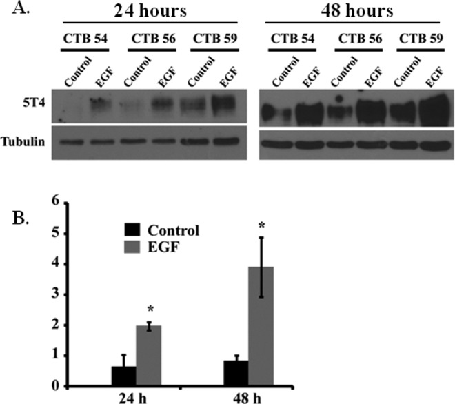
Methods: Trophoblast glycoprotein expression was analyzed by real-time reverse transcription-polymerase chain reaction (RT-PCR), Western blot, and immunohistochemistry. Regulation of 5T4 in cytotrophoblast cells was examined under either differentiating conditions of epidermal growth factor or under varying oxygen conditions. Microvesicles and exosomes were purified from supernatant of cultured and perfused placentas.

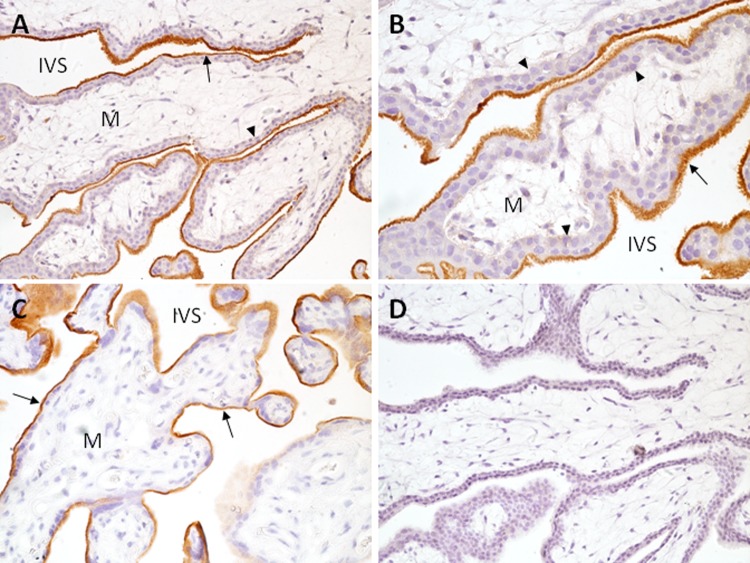
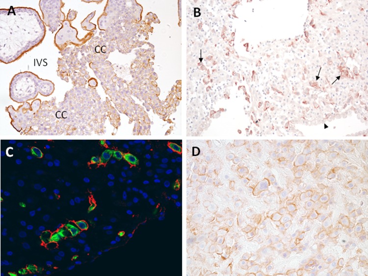
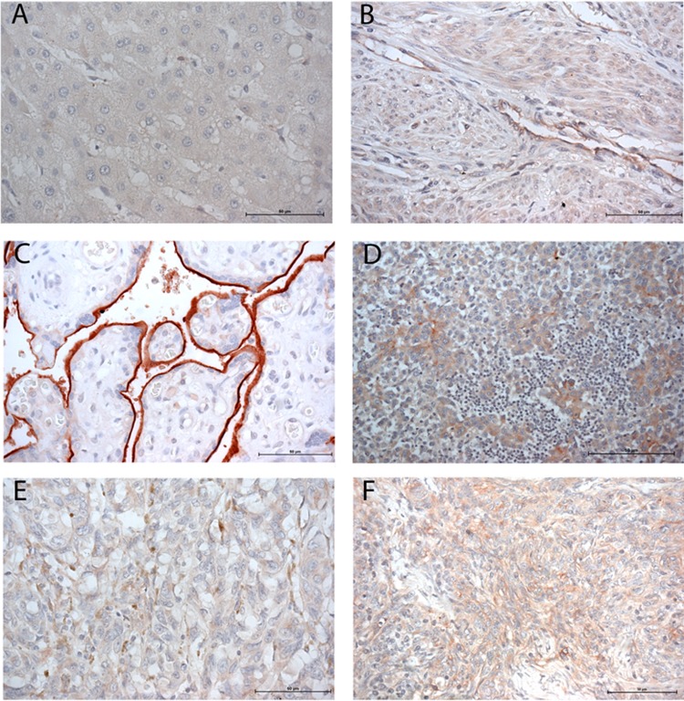
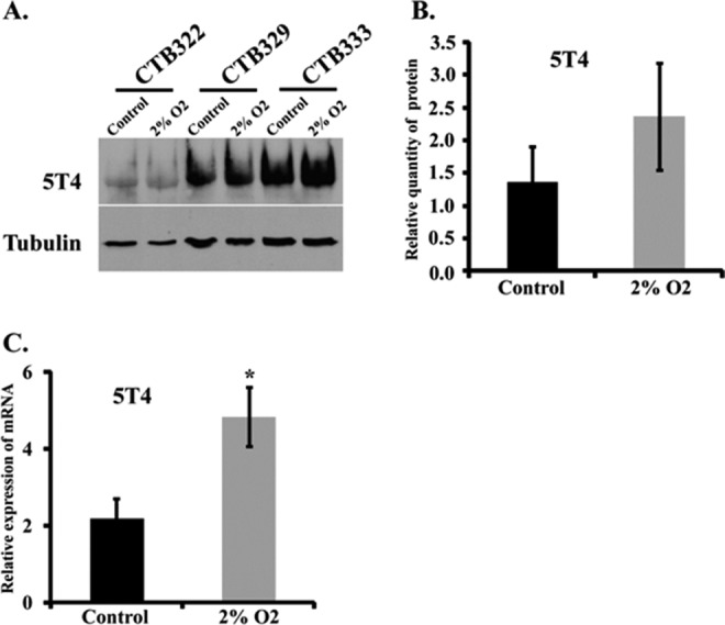
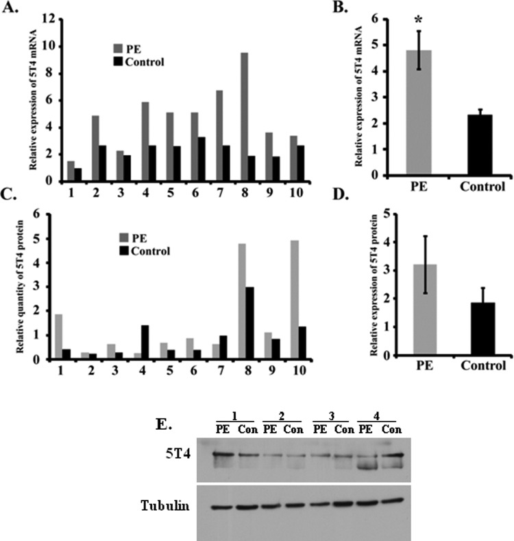
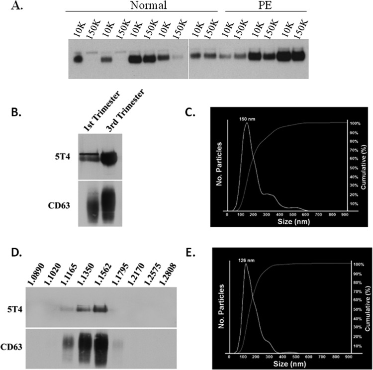
Results: Trophoblast glycoprotein expression was prominent at the microvillus surface of syncytiotrophoblast and on the extravillous trophoblast cells, with minimal expression in undifferentiated cytotrophoblasts and normal tissues. Trophoblast glycoprotein expression was elevated in malignant tumors. In cytotrophoblasts, 5T4 was induced by in vitro differentiation, and its messenger RNA (mRNA) was increased under conditions of low oxygen. PE placentas expressed higher 5T4 mRNA than matched control placentas. Trophoblast glycoprotein was prominent within shed placental microvesicles and exosomes.

Conclusion: Given the potential functional and known immunological importance of 5T4 in cancer, these studies reveal a class of proteins that may influence placental development and/or sensitize the maternal immune system. In extravillous trophoblasts, 5T4 may function in epithelial-to-mesenchymal transition during placentation. The role of syncytiotrophoblast 5T4 is unknown, but its abundance in shed syncytial vesicles may signify route of sensitization of the maternal immune system.

Keywords: 5T4; cancer; exosomes; microvesicles; placenta; trophoblast.


Conflict of interest statement

Declaration of Conflicting Interests: The author(s) declared no potential conflicts of interest with respect to the research, authorship, and/or publication of this article.

Figures

Figure 1.
Figure 1.
Expression of 5T4 in first trimester and term chorionic villi. First trimester (A, B, D) and term (C) tissue sections were incubated with antihuman 5T4 antibody (A-C) or IgG (D) for immunohistochemistry as described in Methods. A, B, Villous placenta showing prominent 5T4 expression (red-brown staining) on microvillus surface of syncytiotrophoblast (arrows); underlying cytotrophoblast cells (arrowheads) and villous mesenchyme (M) lack obvious 5T4 expression. C, Term villous placenta showing strong immunoreactivity at the apical surface of the syncytiotrophoblast (arrow). A, C, and D, 200× magnification; B, 400× magnification; IVS indicates intervillous space; 5T4, trophoblast glycoprotein.
Figure 2.
Figure 2.
Expression of 5T4 in first trimester and term extravillous trophoblast cells. A. Villous placental tissue showing extravillous trophoblast cell columns (CC). B, First trimester decidual tissue containing prominently stained interstitial trophoblast cells (arrows); decidual cells and luminal epithelial cells (arrowhead) are unstained. C, interstitial trophoblast cells are costained for 5T4 (red) and cytokeratin 7 (green), demonstrating surface-associated 5T4 on these cells. D, Term basal plate placenta; extravillous trophoblast cells express 5T4; 5T4 indicates trophoblast glycoprotein.
Figure 3.
Figure 3.
Placental expression of 5T4 across gestation. A, Whole placenta lysate was examined by Western blot analysis using anti-5T4 antibody, or anti-α/β-tubulin as a loading control. B, Semiquantitative analysis of 5T4 in first trimester (n = 3), second trimester (n = 4), and term (n = 6) placenta by scanning densitometry. Samples were normalized to tubulin and analyzed by 1-way analysis of variance (ANOVA). C, Western blot analysis of purified term cytotrophoblast cells (CTB), first trimester trophoblast cell lines (SW71 and HTR8/SVneo), and choriocarcinoma cells (BeWo and JEG-3). HEK-293 cells were used as a negative control. 5T4 indicates trophoblast glycoprotein.
Figure 4.
Figure 4.
Immunoreactive 5T4 in normal and malignant tissues. A commercially obtained tissue microarray was screened for expression of 5T4 across normal and malignant tissues. Tissues shown are liver (A), uterus (B), placenta (C), breast carcinoma (D), thymoma (E), and melanoma (F).
Figure 5.
Figure 5.
EGF on 5T4 expression in primary trophoblast cells. A, Western blot analysis of 3 different samples of purified trophoblast cells cultured in the presence or absence of EGF for 24 or 48 hours. CTB (number) represents the deidentification code used to denote individual samples. Tubulin was used as a loading control. B, Semiquantitative analysis of 5T4 protein expression. Asterisks denote significant differences as compared to controls within time points (P < .05). Data were analyzed by 2-tailed t test; CTB indicates cytotrophoblast; EGF, epidermal growth factor.
Figure 6.
Figure 6.
Low oxygen increases 5T4 messenger RNA (mRNA) expression. A. Western blot analysis of 3 different samples of purified trophoblast cells cultured under ambient (control) or low-oxygen (2% O2) conditions. CTB (number) represents the deidentification code used to denote individual samples. B and C, Semiquantitative analysis of the effect of low oxygen on 5T4 expression on protein (B) and mRNA (C) expression; CTB indicates cytotrophoblast.
Figure 7.
Figure 7.
5T4 messenger RNA (mRNA) transcripts are elevated in preeclampsia. 5T4 protein and mRNA expression were examined in 10 preeclamptic (PE) and 10 gestational age- and delivery-matched healthy control placentas by reverse transcription-polymerase chain reaction (RT-PCR) and Western blot, respectively. Individual (A and C) and mean (B and D) normalized values of mRNA (A and B) and protein (C and D) are shown among samples. Asterisk indicates statistically significant difference as determined by 2-tailed t test. E, Representative Western blot; 5T4 indicates trophoblast glycoprotein.
Figure 8.
Figure 8.
Trophoblast glycoprotein (5T4) is present within exosomes isolated from first trimester and term placental explant culture. A, Placentas from normal healthy pregnancies or pregnancies complicated by preeclampsia (PE) were perfused ex vivo, and perfusates were subjected to centrifugation at 10 000g (10K) or 150 000g (150K). Resulting pellets were analyzed by Western blot for 5T4 expression. B-D, Placental explants were cultured for 24 hours in exosome-free medium, and supernatant subjected to sequentially increasing centrifugation. Pellets were examined by Western blot analysis (B) and nanoparticle tracking analysis (C). D and E, Pellets were further subjected to sucrose gradient centrifugation and similarly examined. Numbers along top of image in D represent densities (g/mL) of each collected fraction.

Similar articles

Cited by

References

    1. Pijnenborg R, Dixon G, Robertson WB, Brosens I. Trophoblast invasion of human decidua from 8-18 weeks of pregnancy. Placenta. 1980;1(1):3–19. - PubMed
    1. Harris LK, Aplin JD. Vascular remodeling and extracellular matrix breakdown in the uterine spiral arteries during pregnancy. Reprod Sci. 2007;14(suppl 8):28–34. - PubMed
    1. Bulmer JN, Innes BA, Levey J, Robson SC, Lash GE. The role of vascular smooth muscle cell apoptosis and migration during uterine spiral artery remodeling in normal human pregnancy. FASEB J. 2012;26(7):2975–2985. - PubMed
    1. Pijnenborg R, Robertson WB, Brosnens I, Dixon G. Review article: trophoblast invasion and the establishment of haemochorial placentation in man and laboratory animals. Placenta. 1981;2(1):71–91. - PubMed
    1. Murray MJ, Lessey BA. Embryo implantation and tumor metastasis: common pathways of invasion and angiogenesis. Semin Reprod Endocrinol. 1999;17(3):275–290. - PubMed

Publication types

Substances

LinkOut - more resources