. 2017 May 8;9(5):156.
doi: 10.3390/toxins9050156.

Histopathological Effects of Bt and TcdA Insecticidal Proteins on the Midgut Epithelium of Western Corn Rootworm Larvae (Diabrotica virgifera virgifera)

Affiliations

Histopathological Effects of Bt and TcdA Insecticidal Proteins on the Midgut Epithelium of Western Corn Rootworm Larvae (Diabrotica virgifera virgifera)

Andrew J Bowling et al. Toxins (Basel). .

Abstract

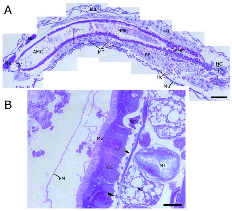
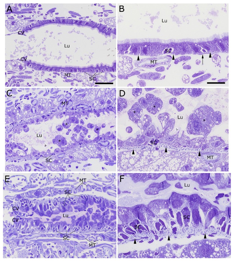
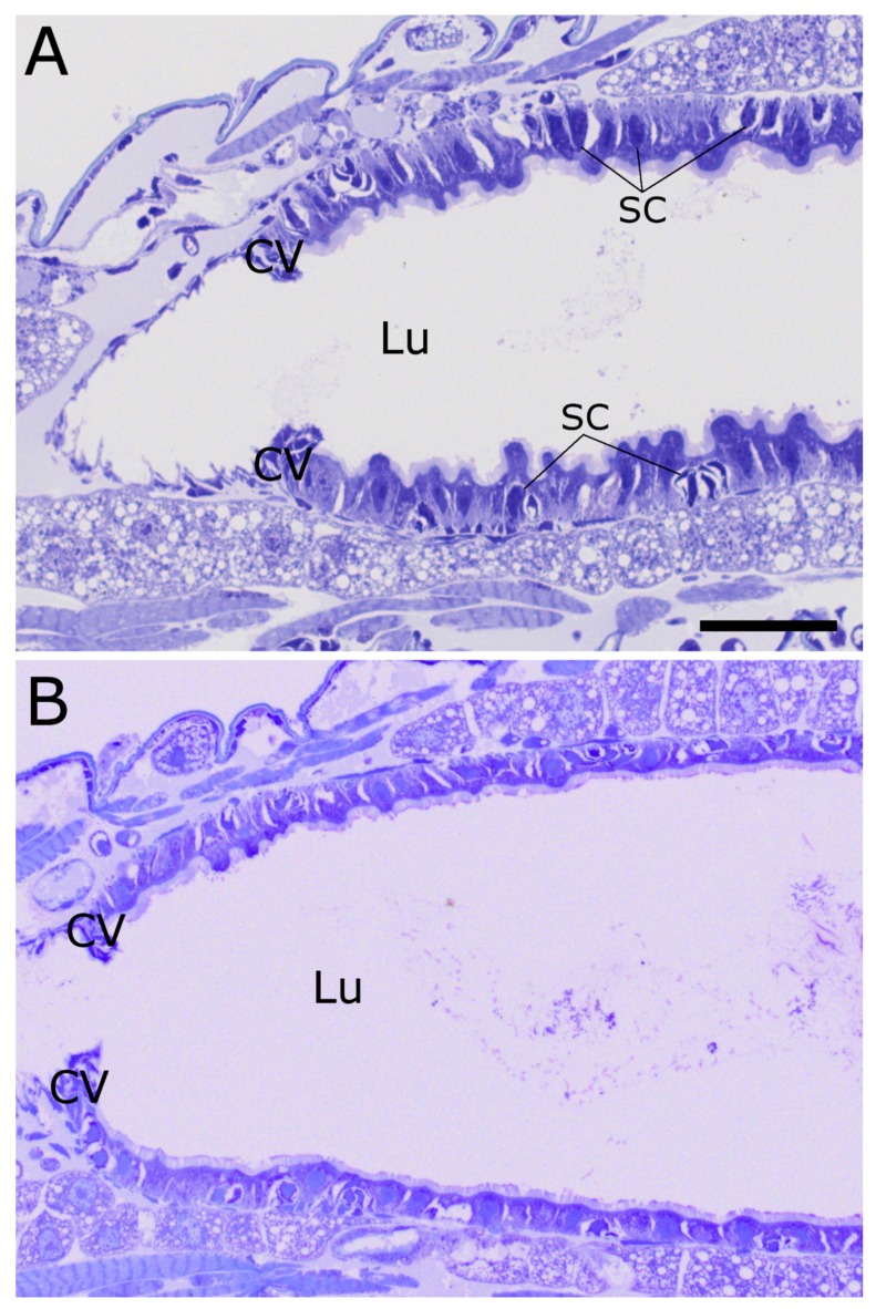
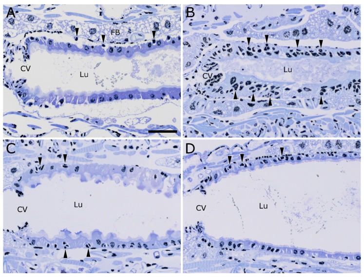
Western corn rootworm (WCR, Diabrotica virgifera virgifera LeConte) is a major corn pest in the United States, causing annual losses of over $1 billion. One approach to protect against crop loss by this insect is the use of transgenic corn hybrids expressing one or more crystal (Cry) proteins derived from Bacillus thuringiensis. Cry34Ab1 and Cry35Ab1 together comprise a binary insecticidal toxin with specific activity against WCR. These proteins have been developed as insect resistance traits in commercialized corn hybrids resistant to WCR feeding damage. Cry34/35Ab1 is a pore forming toxin, but the specific effects of Cry34/35Ab1 on WCR cells and tissues have not been well characterized microscopically, and the overall histopathology is poorly understood. Using high-resolution resin-based histopathology methods, the effects of Cry34/35Ab1 as well as Cry3Aa1, Cry6Aa1, and the Photorhabdus toxin complex protein TcdA have been directly visualized and documented. Clear symptoms of intoxication were observed for all insecticidal proteins tested, including swelling and sloughing of enterocytes, constriction of midgut circular muscles, stem cell activation, and obstruction of the midgut lumen. These data demonstrate the effects of these insecticidal proteins on WCR midgut cells, and the collective response of the midgut to intoxication. Taken together, these results advance our understanding of the insect cell biology and pathology of these insecticidal proteins, which should further the field of insect resistance traits and corn rootworm management.

Keywords: Bacillus thuringiensis; Cry34Ab1; Cry35Ab1; Diabrotica virgifera virgifera; histopathology; western corn rootworm.


Conflict of interest statement

The authors declare no conflict of interest.

Figures

Figure 1
Figure 1
Longitudinal section of western corn rootworm (WCR) showing major anatomical features. (A) WCR larvae have a simple “tube within a tube” morphology. The AMG (arrowheads) is composed of a single smooth layer of columnar cells bearing an apical brush border of microvilli. The MMG and PMG regions are characterized by folds and ridges of the gut epithelium. Between the foregut and the anterior midgut is a valve-like structure called the cardiac valve. Fat bodies (FB) and Malpighian tubules (MT) surround the alimentary canal; (B) Single layers of peritrophic matrix material are visible in the anterior midgut, but become multi-lamellar in the median and posterior midgut regions. Towards the basal region of the columnar cell layer are pockets of stem cells (SC). The adipocytes (Ac) of WCR larvae contain a large number of oil bodies, which appear very translucent by this preparation method. Malpighian tubules, here seen in cross section, can be seen to have a thick layer of microvilli which appear similar but distinct from those lining the alimentary canal. Surrounding the columnar cells are two layers of muscle fibers, both circular (arrowheads) and longitudinal (arrows). (AMG = anterior midgut; MMG = median midgut; PMG = posterior midgut; CV = cardiac valve; HG = hindgut; Ne = neural tissue; Mu = muscle fibers; Mv = microvilli; CC = columnar cell; PM = peritrophic matrix; SC = stem cells; MT = Malpighian tubules; FB = fat body; Ac = adipocyte; scale bar A = 100 µM, B = 10 µm).
Figure 2
Figure 2
Anterior midgut region of 2nd instar WCR larvae fed Cry34/35Ab1 proteins or buffer. (A,B) Untreated larvae display a normal midgut epithelial layer, with a smooth layer of columnar cells and an open lumen. Circular muscles fibers (arrowheads) are evenly spaced around the midgut, just exterior to the basement membrane. Longitudinal muscle fibers (arrow) lie just external to the circular muscle fibers. (C,D) After feeding on diet treated with Cry34/Cry35Ab1 for 24 h, the columnar cells of the AMG show extensive blebbing and sloughing toward the lumen. The apical brush border is no longer continuous, remaining regions are disrupted and reduced, and cell debris is visible in the lumen (asterisks). The stem cell pockets appear relatively unchanged compared to untreated specimens. The bands of circular muscles (arrowheads) surrounding the alimentary canal appear closer together, possibly indicating that they have contracted. (E,F) At 48 h post-feeding, the lumen of the AMG is nearly completely occluded with cell remnants and swollen columnar cells. The circular muscles (arrowheads) appear to be piling up on each other, possibly as a result of continued intense contractions. The midgut stem cell pockets are also much larger in size, and the cardiac valve appears to have thickened. (CV = cardiac valve; Lu = gut lumen; SC = stem cells; MT = Malpighian tubules; arrowheads = circular muscles; scale bar (A,C,E) = 50 µm; (B,D,F) = 20 µm).
Figure 3
Figure 3
Anterior midgut of WCR larvae fed either Cry34Ab1 or Cry35Ab1 alone at 48 h. (A) Ingestion of Cry34Ab1 alone appears to cause some swelling of the apical portion of the columnar cells into the lumen, but very little actual bursting of the cells. The microvilli are still relatively intact and contiguous, but without the obvious cell bursting and/or microvilli shedding seen with the toxin pair, leading to a relatively open lumen. The stem cell pockets appear somewhat enlarged, although not to the extent seen with the toxin pair. The cardiac valve does appear slightly thickened, but the circular muscle bands do not appear heavily contracted; (B) The ingestion of Cry35Ab1 alone caused very little disturbance to the morphology of the midgut cells, and had very little impact on the alimentary canal as a whole. (SC = stem cells; CV = cardiac valve; Lu = gut lumen; scale bar = 50 µm).
Figure 4
Figure 4
Activation and differentiation of midgut epithelial stem cells following intoxication with Cry34/35Ab1 proteins. Anti-histone H3 immunolabeling (black) on toluidine blue-stained longisections of WCR larvae. (A) An untreated WCR larva, showing the normal distribution of cells/nuclei in the midgut epithelium. The midgut epithelium normally consists of a single layer of cells, except for small pockets of small stem cells located on the basal side of the columnar cell layer (arrowheads); (B) After feeding on Cry34/35Ab1 for 48 h, multiple layers of cells/nuclei can be seen along the anterior midgut (e.g., arrowheads); (C) Larva fed Cry34Ab1 only, showing a single layer of epithelial cells with slightly ruffled apical surfaces, but with apparently non-activated stem cell pockets (arrowheads); (D) Larvae fed Cry35Ab1 only appear very similar to the untreated, showing very little perturbation of the epithelial cells and no obvious activation of the stem cell pockets (arrowheads). (CV = cardiac valve; Lu = gut lumen; scale bar = 50 µm).
Figure 5
Figure 5
AMG of WCR larvae fed with various insecticidal protein toxins, after 48 h of continuous exposure. (A) Ingestion of Cry3Aa induces damage similar to that induced by Cry34/35Ab1, primarily, a massive lysis and shedding of AMG columnar cells (asterisks), stem cell activation and growth, contraction of circular muscle bands (arrows), and occlusion of the midgut lumen by cell debris; (B) Ingestion of Cry 6Aa results in damage similar to that induced by Cry34/35Ab1, including overall midgut cell disorder, columnar cell blebbing and lysis, contraction of the circular muscle bands (arrows), and stem cell activation. The lumen is completely full of an apparently non-cellular material, possibly columnar cell lysates and/or secretions. The stem cells appear to be activated and have begun differentiating into replacement enterocytes; (C) TcdA: the stem cells in the AMG appear to have been stimulated and are in advanced stages of columnar cell replacement. Remnants of the columnar cells can still be seen lining the gut lumen (asterisks). The apical microvilli of the columnar cell remnants appear to be relatively intact (arrowheads). The circular muscles appear contracted (arrows), but the lumen does not appear to be entirely occluded. (CV = cardiac valve; SC = stem cells; Lu = gut lumen; MT = Malpighian tubules; scale bar = 50 µm).

Similar articles

Cited by

References

    1. Gray M.E., Sappington T.W., Miller N.J., Moeser J., Bohn M.O. Adaptation and invasiveness of western corn rootworm: Intensifying research on a worsening pest. Annu. Rev. Entomol. 2009;54:303–321. doi: 10.1146/annurev.ento.54.110807.090434. - DOI - PubMed
    1. Metcalf R.L. Foreword. In: Krysan J.L., Miller T.A., editors. Methods for the Study of Pest Diabrotica. Springer; New Youk, NY, USA: 1986. pp. vii–xv.
    1. Narva K.E., Siegfried B.D., Storer N.P. Transgenic approaches to western corn rootworm control. Adv. Biochem. Eng. Biotechnol. 2013;136:135–162. - PubMed
    1. Environmental Protection Agency (EPA) Biopesticides Registration Action Document. Bacillus thuringiensis Cry3Bb1 Protein and the Genetic Material Necessary for Its Production (Vector PV-ZMIR13L) in MON 863 Corn (OECD Unique Identifier: MON-ØØ863-5) [(accessed on 6 May 2017)]; Available online: https://www3.epa.gov/pesticides/chem_search/reg_actions/registration/dec....
    1. Environmental Protection Agency (EPA) Biopesticides Registration Action Document. Modified Cry3A Protein and the Genetic Material Necessary for its Production (Via Elements of pZM26) in Event MIR604 Corn SYN-IR604-8. [(accessed on 6 May 2017)]; Available online: http://www3.epa.gov/pesticides/chem_search/reg_actions/pip/mcry3a-brad.pdf.

LinkOut - more resources