. 2017 Jul;13(7):730-736.
doi: 10.1038/nchembio.2376. Epub 2017 May 8.

Plasticity, dynamics, and inhibition of emerging tetracycline resistance enzymes

Affiliations

Plasticity, dynamics, and inhibition of emerging tetracycline resistance enzymes

Jooyoung Park et al. Nat Chem Biol. 2017 Jul.

Abstract

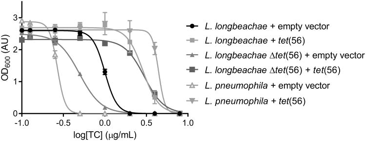
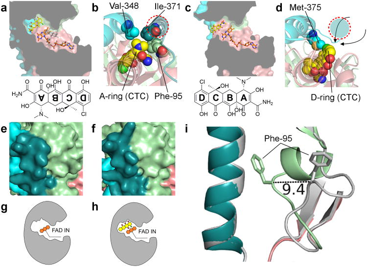
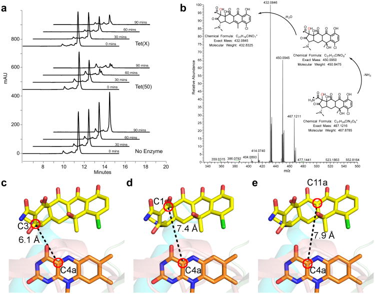
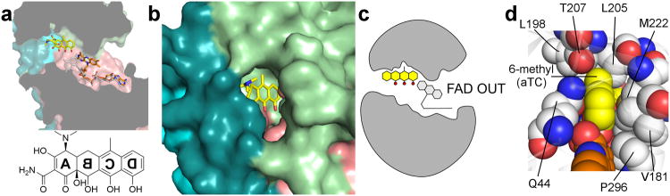
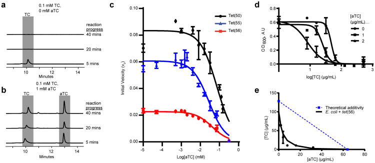
Although tetracyclines are an important class of antibiotics for use in agriculture and the clinic, their efficacy is threatened by increasing resistance. Resistance to tetracyclines can occur through efflux, ribosomal protection, or enzymatic inactivation. Surprisingly, tetracycline enzymatic inactivation has remained largely unexplored, despite providing the distinct advantage of antibiotic clearance. The tetracycline destructases are a recently discovered family of tetracycline-inactivating flavoenzymes from pathogens and soil metagenomes that have a high potential for broad dissemination. Here, we show that tetracycline destructases accommodate tetracycline-class antibiotics in diverse and novel orientations for catalysis, and antibiotic binding drives unprecedented structural dynamics facilitating tetracycline inactivation. We identify a key inhibitor binding mode that locks the flavin adenine dinucleotide cofactor in an inactive state, functionally rescuing tetracycline activity. Our results reveal the potential of a new tetracycline and tetracycline destructase inhibitor combination therapy strategy to overcome resistance by enzymatic inactivation and restore the use of an important class of antibiotics.


Figures

Figure 1
Figure 1. Dose-response curve showing the effect of tetracycline on growth of Legionella strains
Deletion of tet(56) from L. longbeachae causes an increase in tetracycline sensitivity. Complementation with a plasmid containing the tet(56) insert rescues the tetracycline resistance phenotype compared to strains bearing the empty-vector control. Furthermore, introduction of the complementing vector into L. pneumophila, which lacks a tet(56) homolog, results in an increase in tetracycline resistance. Data are represented as mean ± s.d. of three technical replicates.
Figure 2
Figure 2. Crystal structures of Tet(50), Tet(51), Tet(55), and Tet(56) reveal a conserved architecture, structural changes that enable substrate loading channel accessibility, and two conformations of the FAD cofactor
(a) Overlay of the Tet(50) monomer A, Tet(50) monomer B, Tet(51), Tet(55), and Tet(56) crystal structures. The FAD-binding domain (salmon), the tetracycline-binding domain (pale green), the first (cyan) and second (deep teal) C-terminal α-helixes, and FAD molecules (orange) are shown. b-d Surface representation of (b) Tet(50) monomer A with the substrate-loading channel closed, (c) Tet(50) monomer B with the substrate-loading channel open, and (d) a previously published structure of Tet(X) with chlortetracycline (yellow) bound – PDB ID 2Y6R. e-g Zoomed in view of (e) the closed substrate-loading channel in Tet(50) monomer A (f) the open substrate-loading channel in Tet(50) monomer B, and (g) the wide open substrate-binding site in Tet(X). (h) The FAD cofactor adopts the IN conformation in Tet(50) monomer A, characterized by a 12.3 Å distance between the C8M and C2B atoms of the FAD molecule (i) The FAD cofactor adopts the OUT conformation in Tet(50) monomer B, characterized by a 5.2 Å distance between the C8M and C2B atoms of the FAD molecule. (j) The IN conformation of FAD allows for substrate catalysis. The OUT conformation of FAD allows for regeneration of the reduced FAD for the next round of catalysis. The green area indicates the substrate-binding site. The pink area indicates the putative NADPH binding site.
Figure 3
Figure 3. Tet(50)+chlortetracycline structure reveals an unexpected mode of binding that drives substrate loading channel closure and FAD conversion
(a) Chlortetracycline binds to Tet(50) in a ∼180° rotated orientation relative to Tet(X)+chlortetracycline, with FAD IN (orange); a model of FAD OUT (grey) is overlaid. (b) The rotated orientation in the Tet(50)+chlortetracycline structure is supported by van der Waals contacts from Val-348 (cyan) and Ile371 (deep teal) of the two C-terminal α-helices in Tet(50) to the dimethylamine group of the A-ring of chlortetracycline. Additionally, Phe-95 from the flexible loop makes contacts with the dimethylamine group and closes off the substrate-binding site. (c) Chlortetracycline binds Tet(X) with the D-ring distal to FAD. The substrate-binding site is widely exposed to bulk solvent. (d) Met-375 from the first C-terminal α-helix in Tet(X) (cyan) makes van der Waals contacts to the D-ring of chlortetracycline. A second C-terminal helix (red dashed circle, colored in deep teal) does not exist in Tet(X), and substrate can potentially enter from various possible directions. (e) Surface representation of Tet(50)+chlortetracycline monomer A. (f) Surface representation of Tet(50)+chlortetracycline monomer B. (g) In Tet(50)+chlortetracycline monomer A, FAD is IN, the loop is closed, and no chlortetracycline is bound. (h) In Tet(50)+chlortetracycline monomer B, FAD is IN, the loop is closed, and chlortetracycline is bound. (i) While the substrate-loading channel is open in Tet(50) monomer B, with FAD OUT, in the absence of chlortetracycline (grey), the flexible loop containing Phe-95 closes over the channel in Tet(50)+chlortetracycline monomer B, with FAD now IN.
Figure 4
Figure 4. Chlortetracycline is degraded by tetracycline destructases despite the unusual binding mode
(a) HPLC chromatograms show the time and enzyme dependent consumption of chlortetracycline. (b) High-resolution MS-MS analysis of the tetracycline destructase reaction with chlortetracycline supports clean conversion to the m/z 467 oxidation product. MS-MS spectrum of the m/z 467 ion from the Tet(55) reaction with proposed fragmentation pathway. c-e The closest reactive carbons to C4a of the FAD cofactor are C3 (c) and C1 (d) of the chlortetracycline A ring, both of which are closer than C11a (e), the hydroxylation site observed in Tet(X) mediated chlortetracycline degradation.
Figure 5
Figure 5. Anhydrotetracycline binds to the active site of Tet(50), trapping FAD in the unproductive OUT conformation
(a) Anhydrotetracycline binds the active site of Tet(50) and traps the FAD cofactor in the unproductive OUT conformation (orange) in monomer B. The IN conformation of FAD from monomer A is superimposed in grey for comparison, and sterically clashes with the D-ring hydroxyl of anhydrotetracycline. (b) Surface representation of Tet(50)+anhydrotetracycline reveals that the substrate-loading channel remains open, which corresponds to FAD locked in the OUT conformation. (c) In Tet(50)+anhydrotetracycline monomer B, FAD is OUT, the loop is open, and anhydrotetracycline is bound (not shown: in monomer A, FAD is IN, the loop is closed, no anhydrotetracycline is bound). (d) Residue Thr-207 in Tet(50) makes van der Waals interactions with the planar 6-methyl group of anhydrotetracycline (aTC) (yellow) in the bound orientation, but would sterically clash with the 6-methyl and 6-hydroxyl groups that branch from the C ring of tetracycline or chlortetracycline if bound in a flipped orientation.
Figure 6
Figure 6. Anhydrotetracycline prevents enzymatic tetracycline degradation, functionally rescuing tetracycline antibiotic activity
(a) Tetracycline (TC) is degraded by Tet(56) in vitro HPLC chromatograms show in vitro reactions with UV detection at 363 nm and separation on a C18 column. (b) TC degradation is attenuated by the addition of an excess of aTC. (c) Dose-dependent inhibition of Tet(50,51,56) activity by anhydrotetracycline. Velocity is determined by measuring tetracycline consumption via change in absorbance at 400 nm. Data are represented as mean ± s.d. of three technical replicates. (d) Dose-response curve showing effect of aTC on sensitivity of Tet(56)-expressing E. coli to TC in liquid culture. Data are represented as mean ± s.e.m. of three technical replicates. (e) TC and aTC synergistically inhibit growth of E. coli expressing Tet(56), FICI = 0.1875. Points show minimum inhibitory concentrations of two drugs in combination. Dashed line indicates the theoretical concentration of additive drug interaction. Data represented as mean ± s.e.m. of three technical replicates.

Comment in

References

    1. Knapp CW, Dolfing J, Ehlert PA, Graham DW. Evidence of increasing antibiotic resistance gene abundances in archived soils since 1940. Environ Sci Technol. 2010;44:580–587. doi: 10.1021/es901221x. - DOI - PubMed
    1. Kinch MS, Patridge E, Plummer M, Hoyer D. An analysis of FDA-approved drugs for infectious disease: antibacterial agents. Drug Discov Today. 2014;19:1283–1287. doi: 10.1016/j.drudis.2014.07.005. - DOI - PubMed
    1. Davies J. Inactivation of antibiotics and the dissemination of resistance genes. Science. 1994;264:375–382. - PubMed
    1. Allen HK, et al. Call of the wild: antibiotic resistance genes in natural environments. Nat Rev Microbiol. 2010;8:251–259. doi: 10.1038/nrmicro2312. - DOI - PubMed
    1. Berendonk TU, et al. Tackling antibiotic resistance: the environmental framework. Nat Rev Microbiol. 2015;13:310–317. doi: 10.1038/nrmicro3439. - DOI - PubMed

online Methods References

    1. Merriam JJ, Mathur R, Maxfield-Boumil R, Isberg RR. Analysis of the Legionella pneumophila fliI gene: intracellular growth of a defined mutant defective for flagellum biosynthesis. Infect Immun. 1997;65:2497–2501. - PMC - PubMed
    1. Feeley JC, et al. Charcoal-yeast extract agar: primary isolation medium for Legionella pneumophila. J Clin Microbiol. 1979;10:437–441. - PMC - PubMed
    1. Berger KH, Isberg RR. Two distinct defects in intracellular growth complemented by a single genetic locus in Legionella pneumophila. Mol Microbiol. 1993;7:7–19. - PubMed
    1. Sexton JA, et al. The Legionella pneumophila PilT homologue DotB exhibits ATPase activity that is critical for intracellular growth. J Bacteriol. 2004;186:1658–1666. - PMC - PubMed
    1. Kabsch W. XDS. Acta Crystallogr D Biol Crystallogr. 2010;66:125–132. doi: 10.1107/s0907444909047337. - DOI - PMC - PubMed

MeSH terms