. 2017 Jun;23(6):768-774.
doi: 10.1038/nm.4329. Epub 2017 May 8.

Decreased alertness due to sleep loss increases pain sensitivity in mice

Affiliations

Decreased alertness due to sleep loss increases pain sensitivity in mice

Chloe Alexandre et al. Nat Med. 2017 Jun.

Abstract

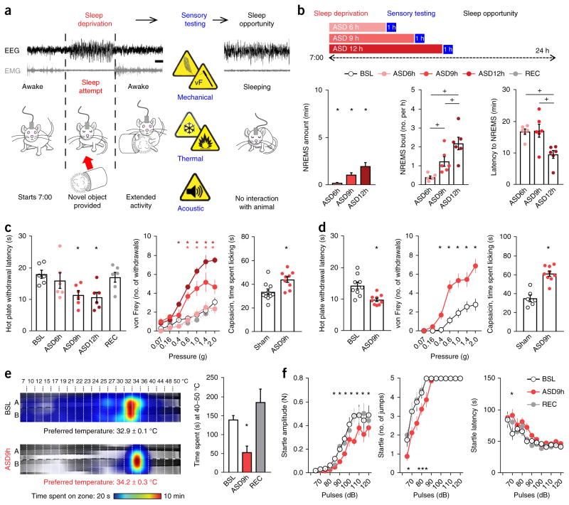
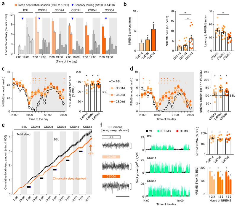
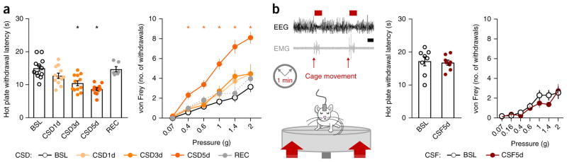
Extended daytime and nighttime activities are major contributors to the growing sleep deficiency epidemic, as is the high prevalence of sleep disorders like insomnia. The consequences of chronic insufficient sleep for health remain uncertain. Sleep quality and duration predict presence of pain the next day in healthy subjects, suggesting that sleep disturbances alone may worsen pain, and experimental sleep deprivation in humans supports this claim. We demonstrate that sleep loss, but not sleep fragmentation, in healthy mice increases sensitivity to noxious stimuli (referred to as 'pain') without general sensory hyper-responsiveness. Moderate daily repeated sleep loss leads to a progressive accumulation of sleep debt and also to exaggerated pain responses, both of which are rescued after restoration of normal sleep. Caffeine and modafinil, two wake-promoting agents that have no analgesic activity in rested mice, immediately normalize pain sensitivity in sleep-deprived animals, without affecting sleep debt. The reversibility of mild sleep-loss-induced pain by wake-promoting agents reveals an unsuspected role for alertness in setting pain sensitivity. Clinically, insufficient or poor-quality sleep may worsen pain and this enhanced pain may be reduced not by analgesics, whose effectiveness is reduced, but by increasing alertness or providing better sleep.


Conflict of interest statement

COMPETING FINANCIAL INTERESTS

The authors declare no competing financial interests.

Figures

Figure 1
Figure 1
Acute sleep deprivation progressively increases sleepiness and sensitivity to pain. (a) Schematic representation of the sleep deprivation protocol: an experimenter continuously monitored EEG and EMG of the mice and placed a novel object in their cage to promote arousal whenever an NREMS entry (sleep attempt) was detected. Sensory testing (in blue) took place immediately after the sleep deprivation period; mice were then allowed to sleep ad libitum (sleep opportunity). Scale bar, 5 s. (b) Top, ASD was carried out for 6, 9 or 12 h starting at 7:00. Left, NREMS amount expressed as percentage of corresponding baseline values. Middle, the number of NREMS entries (bouts lasting at least 5 s). Right, latency to entering NREMS upon return of the mice to their home cage (n = 6 mice). BSL, assessment at baseline; REC, assessment after a night of recovery sleep. (c) Sensory assessment in male mice. Left, latency to withdrawal from contact with a 52 °C hot plate (n = 6 mice). Middle, mechanical sensitivity measured as the number of brisk withdrawals from ten stimulations with von Frey filaments of different forces applied to the plantar surface of the hindpaw (n = 6 mice). Right, nocifensive response (time spent licking) after intraplantar injection of capsaicin in male mice (1 μg diluted in 20 μl of saline with 1% DMSO; n = 10 sham-treated mice and 9 ASD mice). (d) Sensory assessment in female mice. Left, latency to withdrawal from contact with a 52 °C hot plate (n = 9 mice). Middle, mechanical sensitivity (n = 9 mice). Right, nocifensive response after intraplantar injection of capsaicin (1 μg diluted in 20 μl; n = 6 sham mice and 9 ASD mice). (e) Left, heat map of the time spent in the zones of a thermal gradient at baseline and after 9 h of ASD and the preferred temperature (n = 6 mice). When sleep deprived, mice preferred a slightly warmer temperature. Right, time spent in the noxious heat range (40–50 °C) at baseline, after 9 h of ASD and after recovery (n = 6 mice). (f) Left, intensity, measured as the force produced by a mouse upon jumping; middle, number of jumps; right, latency to jumping. Mice underwent the acoustic startle reflex test at baseline, after 9 h of ASD and after recovery (n = 8 mice). All data are presented as means ± s.e.m. Circles overlaid upon the bar in histograms represent data from each individual animal. *P < 0.05, in comparison to baseline; +P < 0.05, in multiple-group comparisons. For complete statistical analyses (post hoc test, within-subjects or inter-subjects comparison, effect size), please refer to Supplementary Table 1.
Figure 2
Figure 2
Chronic sleep deprivation progressively increases sleepiness. (a) Locomotor activity of mice (3-h intervals) recorded with telemetry throughout CSD. On each day of CSD (CSD1d–CSD5d), mice underwent sleep deprivation (7:00–13:00; orange shading) followed by sleep opportunity periods (14:00–7:00, 17 h total; gray). Sensory testing (13:00; blue triangle) was performed for 1 h at baseline, CSD1d, CSD3d and CSD5d. Periods of darkness (19:00–7:00) are represented as boxes shaded in light gray. (b) Left, NREMS amount expressed as percentage of corresponding baseline values. Middle, number of NREMS entries during sleep deprivation sessions. Right, latency to NREMS onset at the beginning of the sleep opportunity period. (c) Hourly amount of NREMS and rebound sleep computed over the entire sleep opportunity period from 14:00 to 7:00, expressed as percentage of the time-matched baseline amount of NREMS. (d) Hourly amount of REMS and rebound sleep computed over the entire sleep opportunity period from 14:00 to 7:00, expressed as percentage of the time-matched baseline amount of REMS. (e) Cumulative time course of the total sleep amount (summed in 1-h bins) at baseline (black) and on CSD5d (orange). The control curve was generated by cumulatively adding the individual curve obtained at baseline six times. The absence of sleep during each 6-h sleep deprivation session is represented by a solid black rectangle. The net estimated total sleep loss at the end of 5 h of CSD was 7.9 ± 1.5 h. (f) Left, example EEGs of NREMS. Middle, time course of SWA (spectral EEG power in the 0.5- to 4.0-Hz range) during the first 2.5 h of sleep opportunity at baseline, CSD1d and CSD5d in one representative mouse. W, wake. Top right, NREMS SWA computed over 5 h (14:00–19:00) expressed as percentage of the 24-h baseline mean. Bottom right, time course of NREMS SWA during the first 3 h of NREMS after sleep deprivation. Horizontal scale bar, 10 s; vertical scale bar, 500 μV. Data are presented as means ± s.e.m. For all panels, n = 11 mice. Circles overlaid upon the bar in histograms represent data from each individual animal. *P < 0.05, in comparison to baseline; +P < 0.05, in multiple-group comparisons. For complete statistical analyses (post hoc test, within-subjects or inter-subjects comparison, effect size), please refer to Supplementary Table 1.
Figure 3
Figure 3
Chronic sleep deprivation but not chronic sleep fragmentation progressively increases pain sensitivity. (a) Left, latency to withdrawal from noxious heat contact (52 °C hot plate). Right, mechanical sensitivity measured as the number of brisk withdrawals from ten stimulations with von Frey filaments of different forces that were applied to the plantar surface of the hindpaw. Mice were tested at baseline, CSD1d, CSD3d, CSD5d and on recovery (n = 12 mice). (b) Left, schematic representation of the CSF protocol and a representative EEG trace of fragmented sleep. A platform below the cage briefly (10–20 ms) moved upward at a random intensity every minute from 7:00 to 19:00 over five consecutive days. Mice were tested for sensory responsiveness at baseline and CSF5d. Middle, latency to withdrawal from heat contact (52 °C hot plate). Right, mechanical sensitivity. n = 8 mice; nonsignificant Student’s t-test. Scale bar, 10 s. Data are presented as means ± s.e.m. Circles overlaid upon the bar in histograms represent data from each individual animal. *P < 0.05, in comparison to baseline. For complete statistical analyses (post hoc test, within-subjects or inter-subjects comparison, effect size), please refer to Supplementary Table 1.
Figure 4
Figure 4
Caffeine and modafinil, but not ibuprofen or morphine, prevent sleep-deprivation-induced hypersensitivity to pain. (a) Left, latency to withdrawal from noxious heat contact (52 °C hot plate). Right, mechanical sensitivity measured as the number of brisk withdrawals from ten stimulations with von Frey filaments of different forces that were applied to the plantar surface of the hindpaw. Mice were sleep-deprived for 9 h (ASD9h) and injected with saline (SAL, i.p.; red), morphine (5 mg per kg bodyweight, s.c.; dark purple), ibuprofen (IBU, 30 mg per kg bodyweight, s.c.; light purple), caffeine (CAFF, 20 mg per kg bodyweight, i.p.; blue) or modafinil (MOD, 45 mg per kg bodyweight, i.p.; green). Morphine and ibuprofen were administered 30 min before the end of the 9-h ASD period, and caffeine and modafinil were injected 2 h before the end of the 9-h ASD period. n = 8 mice per group. (b) Withdrawal latencies to noxious heat (expressed as percentage of the baseline) of mice treated with saline or morphine (5 mg per kg bodyweight, s.c.) after ad libitum undisturbed sleep and 9 h of sleep deprivation (n = 8 mice per group). (c) Left, heat map of the time spent in the zones of a thermal gradient showing thermal place preference and the calculated preferred temperature. Right, time spent in the noxious heat range (40–50 °C). Sleep-deprived mice were injected with either caffeine (20 mg per kg bodyweight, i.p.) or saline (i.p.) 2 h before the end of the 9-h ASD period (n = 6 mice per group). (d) Mechanical sensitivity in naive animals (ad libitum sleep) 2 h after caffeine (20 mg per kg bodyweight, i.p.), modafinil (45 mg per kg bodyweight, i.p.) or saline (i.p.) injection (n = 13 SAL-, 7 CAFF- and 8 MOD-injected mice). (e) Effects of caffeine, modafinil or ibuprofen on hypersensitivity to mechanically induced pain resulting from peripheral inflammation. Thresholds were assessed at baseline (pre-CFA), the day after intraplantar injection of CFA and 2 h after administration of saline, caffeine or modafinil (n = 8 SAL-, 8 CAFF- and 7 MOD-injected mice) and 30 min after administration of ibuprofen (n = 7 mice). (f) Left, withdrawal latency to contact with heat. Right, mechanical sensitivity. Mice undergoing CSD (6 h daily) were injected with saline (i.p.) or caffeine (20 mg per kg bodyweight, i.p.) on day 4 of CSD (n = 6 mice). (g) Latency to NREMS onset at the beginning of the sleep opportunity period (left) and NREMS rebound (right) following administration of saline on CSD4d and caffeine (20 mg per kg bodyweight, i.p.) on CSD5d computed over the entire sleep opportunity period from 14:00 to 7:00, expressed as percentage of corresponding baseline values (n = 5 mice). Data are presented as means ± s.e.m. Circles overlaid upon the bar in histograms represent data from each individual animal. *P < 0.05, in comparison to baseline; +P < 0.05, in multiple-group comparisons. For complete statistical analyses (post hoc test, within-subjects or inter-subjects comparison, effect size), please refer to Supplementary Table 1.

Comment in

  • Say good night to your pain.
    Lehtinen MK. Lehtinen MK. Sci Transl Med. 2017 May 31;9(392):eaan4298. doi: 10.1126/scitranslmed.aan4298. Sci Transl Med. 2017. PMID: 28566427

References

    1. Van den Bulck J. Television viewing, computer game playing, and Internet use and self-reported time to bed and time out of bed in secondary-school children. Sleep. 2004;27:101–104. - PubMed
    1. Fossum IN, Nordnes LT, Storemark SS, Bjorvatn B, Pallesen S. The association between use of electronic media in bed before going to sleep and insomnia symptoms, daytime sleepiness, morningness, and chronotype. Behav Sleep Med. 2014;12:343–357. - PubMed
    1. Luyster FS, Strollo PJ, Jr, Zee PC, Walsh JK. Sleep: a health imperative. Sleep. 2012;35:727–734. - PMC - PubMed
    1. Edwards RR, Almeida DM, Klick B, Haythornthwaite JA, Smith MT. Duration of sleep contributes to next-day pain report in the general population. Pain. 2008;137:202–207. - PMC - PubMed
    1. Campbell CM, et al. Self-reported sleep duration associated with distraction analgesia, hyperemia, and secondary hyperalgesia in the heat–capsaicin nociceptive model. Eur J Pain. 2011;15:561–567. - PMC - PubMed

MeSH terms