. 2017 Jul;96(8):909-916.
doi: 10.1177/0022034517707513. Epub 2017 May 8.

Contribution of the PDL to Osteotomy Repair and Implant Osseointegration

Affiliations

Contribution of the PDL to Osteotomy Repair and Implant Osseointegration

X Pei et al. J Dent Res. 2017 Jul.

Abstract

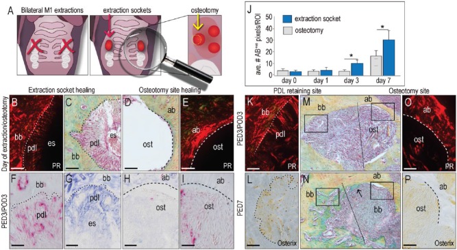
Our objective was to clarify the fate of the periodontal ligament (PDL) retained in the socket after tooth extraction, then determine if this tissue contributed to the osseointegration of "immediate" implants placed in these fresh extraction sockets. Mice underwent maxillary first molar extraction, the residual PDL was removed by an osteotomy, and titanium implants were placed. The osteotomy was created in such a way that the palatal surface was devoid of PDL remnants while the buccal, mesial, and distal surfaces retained PDL fibers. At multiple time points after surgery, tissues were analyzed using a battery of molecular, cellular, and histomorphometrical assays. We found that PDL remnants mineralized and directly contributed to new bone formation in the extraction site. Compared with regions of an extraction site where the PDL was removed by osteotomy, regions that retained PDL fibers had produced significantly more new bone. Around immediate implants, the retained PDL remnants directly contributed to new bone formation and osseointegration. Thus, we conclude that PDL remnants are inherently osteogenic, and if the tissue is healthy, it is reasonable to conclude that curetting out an extraction socket prior to immediate implant placement should be avoided. This recommendation aligns with contemporary trends toward minimally invasive surgical manipulations of the extraction socket prior to immediate implant placement.

Keywords: bone; extraction socket; histology; immediate implant; periostin; regeneration.


Conflict of interest statement

The authors declare no potential conflicts of interest with respect to the authorship and/or publication of this article.

Figures

Figure 1.
Figure 1.
Molecular and cellular events characterizing extraction socket healing. (A) Representative transverse tissue section through the mouse maxilla stained with aniline blue, illustrating the position of the first molar (M1), its periodontal ligament (PDL), and surrounding alveolar bone. Black bracket indicates the cementum. (B) Aniline blue staining of the extraction socket; in all panels, the asterisks identify PDL remnants attached to the bundle bone. (C) On postextraction day (PED) 1, sagittal tissue section immunostained with periostin. (D) On PED2, tartrate-resistant acid phosphatase (TRAP) and alkaline phosphatase (ALP) activate in the socket. (E) On PED3, the residual PDL expresses osterix. (F) Pentachrome staining of sagittal sections through the extraction socket on PED1, (G) PED4, (H) PED7, and (I) PED14. ab, alveolar bone; bb, bundle bone; c, cementum; d, dentin; es, extraction socket; pdl, periodontal ligament. Scale bars = 100 μm.
Figure 2.
Figure 2.
The periodontal ligament (PDL) retained in the extraction socket undergoes mineralization. (A) Representative sagittal tissue section stained with pentachrome, illustrating the intact M1 PDL. (B) Adjacent tissue section stained with picrosirius red to identify the organization of collagen in the PDL. (C) Periostin immunostaining identifies the intact PDL. On postextraction day (PED) 1, (D) pentachrome staining of the PDL and (E) picrosirius red shows the change in collagen organization after tooth extraction. (F) PDL remnants retain the periostin-positive status. On PED7, the structure of the PDL is notably altered as shown by (G) pentachrome staining. (H) PDL remnants still express periostin; in addition, (I) cells in the PDL remnants strongly express osterix. On PED14, (J) pentachrome, (K) picrosirius red staining, and (L) osterix immunostaining do not differentiate between the mineralized PDL remnants and adjacent alveolar bone. Asterisks indicate PDL remnants. ab, alveolar bone; c, cementum; d, dentin. Scale bars = 50 μm.
Figure 3.
Figure 3.
Periodontal ligament (PDL) remnants contribute to new bone formation. (A) Schematic of the experimental design. On the day of extraction, a representative transverse tissue section stained with (B) picrosirius red to detect the collagenous PDL remnants, and an adjacent section stained with (C) pentachrome staining shows the relationship between PDL remnants the underlying bundle bone after tooth extraction. (D) Removal of the PDL by osteotomy site preparation leaves the alveolar bone smooth and devoid of a (E) collagenous PDL. In all panels, the dotted white line indicates the demarcation between alveolar bone and PDL remnants; the dashed white line indicates the osteotomy edge. (F) Tartrate-resistant acid phosphatase (TRAP) staining and (G) alkaline phosphatase (ALP) activity in the extraction socket on postextraction day (PED) 3. (H) TRAP staining and (I) ALP activity in the osteotomy on postosteotomy day 3 (POD3). (J) Quantification of new bone formation in an extraction socket versus an osteotomy. (M, N) In a single extraction socket, an osteotomy was made, which specifically removed alveolar bone on the palatal aspect but retained the PDL on the mesial, distal, and buccal aspects. Three days later, picrosirius red staining identifies regions (K) with and (O) without PDL remnants. (L, P) The same type of site, evaluated on day 7. Asterisks indicate P < 0.05. ab, alveolar bone; bb, bundle bone; es, extraction socket; ost, osteotomy; pdl, periodontal ligament; PR, picrosirius red staining. Scale bars = 50 μm.
Figure 4.
Figure 4.
Periodontal ligament (PDL) remnants contribute directly to implant osseointegration. (A) Schematic of the experimental design. (B) Schematic of the orientation of the implant in the palatal root and plane of section used to analyze regions of the implant in direct contact with bone, as well as in regions with an intervening PDL. (C) On the day of extraction, a representative transverse tissue section stained with pentachrome in a region with an intervening PDL and (D) a region with direct bone/implant contact. Terminal deoxynucleotidyl transferase dUTP nick end labeling (TUNEL)/4′,6-diamidino-2-phenylindole (DAPI) staining of (E) an intervening PDL region and (F) a direct bone/implant contact region. On postimplant day (PID) 3, tartrate-resistant acid phosphatase (TRAP) staining of a region with (G) an intervening PDL and (H) one with direct bone/implant contact. (I, J) Equivalent regions, stained to detect alkaline phosphatase (ALP) activity. On PID14, (K) bone is in direct contact with the implant in regions that previously had an intervening PDL. (L) In regions that had previously had a direct contact between bone and implant now had a small fibrous envelope. ab, alveolar bone; bb, bundle bone; BIC, bone-implant contact; im, implant; pdl, periodontal ligament.

References

    1. Ahn JJ, Shin HI. 2008. Bone tissue formation in extraction sockets from sites with advanced periodontal disease: a histomorphometric study in humans. Int J Oral Maxillofac Implants. 23(6):1133–1138. - PubMed
    1. Albrektsson T, Branemark PI, Hansson HA, Lindstrom J. 1981. Osseointegrated titanium implants. Requirements for ensuring a long-lasting, direct bone-to-implant anchorage in man. Acta Orthop Scand. 52(2):155–170. - PubMed
    1. Araújo MG, Lindhe J. 2005. Dimensional ridge alterations following tooth extraction: an experimental study in the dog. J Clin Periodontol. 32(2):212–218. - PubMed
    1. Barzilay I, Graser GN, Iranpour B, Proskin HM. 1996. Immediate implantation of pure titanium implants into extraction sockets of Macaca fascicularis. Part I: Clinical and radiographic assessment. Int J Oral Maxillofac Implants. 11(3):299–310. - PubMed
    1. Becker W. 2006. Immediate implant placement: treatment planning and surgical steps for successful outcomes. Br Dent J. 201(4):199–205. - PubMed

LinkOut - more resources