. 2017 Nov 17;8(8):1602-1617.
doi: 10.1080/21505594.2017.1326438. Epub 2017 Jun 1.

S. oralis activates the Efg1 filamentation pathway in C. albicans to promote cross-kingdom interactions and mucosal biofilms

Affiliations

S. oralis activates the Efg1 filamentation pathway in C. albicans to promote cross-kingdom interactions and mucosal biofilms

Hongbin Xu et al. Virulence. .

Abstract

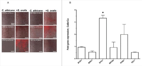
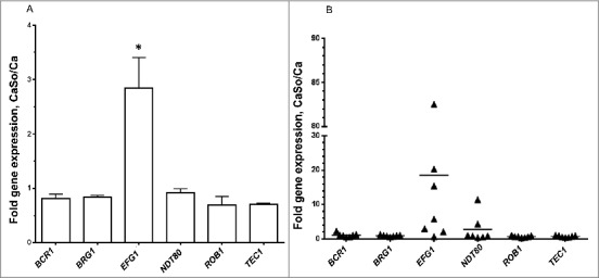
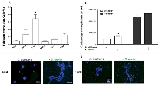
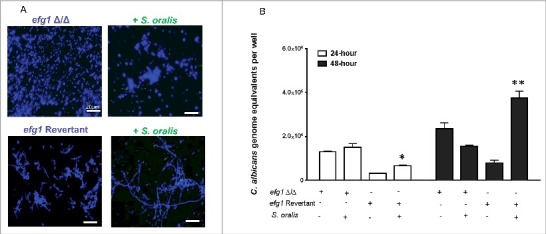
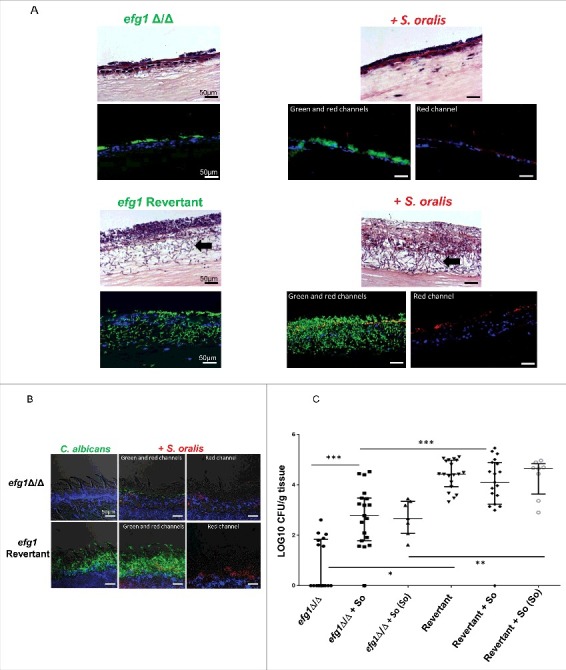
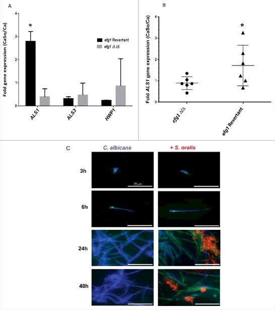
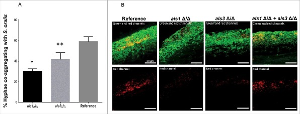
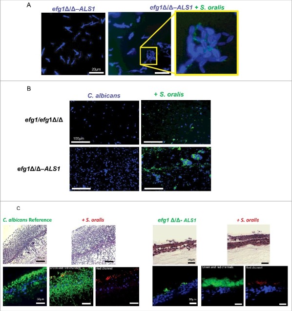
Candida albicans and Streptococcus oralis are ubiquitous oral commensal organisms. Under host-permissive conditions these organisms can form hypervirulent mucosal biofilms. C. albicans biofilm formation is controlled by 6 master transcriptional regulators: Bcr1, Brg1, Efg1, Tec1, Ndt80, and Rob1. The objective of this work was to test whether any of these regulators play a role in cross-kingdom interactions between C. albicans and S. oralis in oral mucosal biofilms, and identify downstream target gene(s) that promote these interactions. Organotypic mucosal constructs and a mouse model of oropharyngeal infection were used to analyze mucosal biofilm growth and fungal gene expression. By screening 6 C. albicans transcription regulator reporter strains we discovered that EFG1 was strongly activated by interaction with S. oralis in late biofilm growth stages. EFG1 gene expression was increased in polymicrobial biofilms on abiotic surfaces, mucosal constructs and tongue tissues of mice infected with both organisms. EFG1 was required for robust Candida-streptococcal biofilm growth in organotypic constructs and mouse oral tissues. S. oralis stimulated C. albicans ALS1 gene expression in an EFG1-dependent manner, and Als1 was identified as a downstream effector of the Efg1 pathway which promoted C. albicans-S. oralis coaggregation interactions in mixed biofilms. We conclude that S. oralis induces an increase in EFG1 expression in C. albicans in late biofilm stages. This in turn increases expression of ALS1, which promotes coaggregation interactions and mucosal biofilm growth. Our work provides novel insights on C. albicans genes which play a role in cross-kingdom interactions with S. oralis in mucosal biofilms.

Keywords: ALS1; Candida; EFG1; Streptococcus; cross-kingdom biofilm; oral mucosa.


Figures

Figure 1.
Figure 1.
S. oralis activates C. albicans EFG1 gene expression in mixed biofilms. (A) C. albicans mCherry transcriptional regulator reporter strains were grown as biofilms on Permanox® plastic chamber slides, with or without S. oralis 34 in RPMI 10%FBS, 10% BHI media for 36 hours and observed under a fluorescence microscope. A representative of 3 experiments is shown. There was a burst of the red fluorescence signal in the EFG1p-mCherry construct growing with S. oralis, suggesting EFG1 transcriptional activation. Bars: 20 μm. (B) Candida gene mRNA levels from biofilms growing under identical conditions as in (A) were analyzed by RT-qPCR. Results represent fold increase gene expression in C. albicans with S. oralis (CaSo) over C. albicans (Ca) alone. Means ± SD are shown from 3 experiments. S. oralis stimulated a significant increase in EFG1 gene transcripts. *p<0.05 in a comparison to other regulator genes.
Figure 2.
Figure 2.
S. oralis increases EFG1 gene expression of C. albicans in organotypic tissue and mouse oral mucosal infection models. (A) Organotypic tissues were infected with C. albicans SC5314 with or without S. oralis 34, for 16h. Candida gene mRNA levels were analyzed by RT-qPCR. Results represent fold increase in gene expression over the single infection group. Means ± SD are shown from twice repeated experiments, with tissue infections set up in duplicate. EFG1 was the only gene that was significantly upregulated by S. oralis. *p<0.05 for a comparison to all other regulators. (B) Mice were infected with C. albicans or C. albicans plus S. oralis 34 for 4 d and Candida gene mRNA levels in tongue samples were analyzed by RT-qPCR. Results represent fold increase gene expression over the single infection group, with 6–7 mice per group. In most mice infected with both organisms, EFG1 transcripts were higher compared with single infection, although variability in the magnitude of this response was noted.
Figure 3.
Figure 3.
S. oralis promotes C. albicans hyphae in synthetic saliva. (A) C. albicans SC5314 (Ca) transcriptional regulator gene mRNA levels in biofilms growing in 6-well polystyrene plates with or without S. oralis 34 (So) for 24h, with SSM as the sole nutrient source. RNA samples were analyzed by RT-qPCR, after 24 h of co-culture. Results represent fold increase gene expression in mixed (CaSo) over C. albicans alone (Ca) biofilms. Means ± SD are shown from 3 experiments. EFG1 transcripts were significantly increased in the presence of S. oralis, whereas transcription of other regulators was either repressed or essentially unaltered. *p<0.05 in comparison with other regulator genes. (B) C. albicans SC5314 (blue) or C. albicans with teal protein expressing S. oralis 34 (green) were cultured on glass slides with SSM, supplemented with or without 10% BHI for 48 hours. Candida cells were stained with Calcoflour White® and cultures observed under a fluorescence microscope. Co-culture with S. oralis promoted hyphal growth and cell-cell aggregation under both conditions. Bars: 20 μm. (C) Fungal biomass expressed as “genome equivalents” of C. albicans SC5314 in biofilms with or without S. oralis 34. Biofilms were grown in 6-well polystyrene plates with SSM as the sole nutrient source for 24 h or 48 h. Genome equivalents were extrapolated by analyzing 18 S rRNA gene copy numbers in each biofilm well with qPCR and comparing to a standard curve. Means ± SD are shown from 3 experiments. Fungal biomass was higher in biofilms with S. oralis consistent with hyphal growth (B), although this effect was small and reached statistical significance only at the 24 h time-point. *p < 0.05, for a comparison to C. albicans alone.
Figure 4.
Figure 4.
Hyphal growth stimulated by S. oralis is dependent on the Efg1 transcriptional regulator. (A) C. albicans efg1 homozygous deletion mutant (efg1Δ/Δ) and efg1 revertant were grown with or without teal protein expressing S. oralis 34 (green) in 10% BHI-supplemented SSM medium, on Permanox® plastic chamber slides, for 48 hours. Candida cells were stained with Calcoflour White® (blue) and cultures were observed under a fluorescence microscope. The revertant strain formed a mix of yeast and short hyphae in SSM, which were elongated when growing with S. oralis. Bars: 20 μm. (B) Fungal biomass expressed as “genome equivalents” of the efg1Δ/Δ mutant and efg1 revertant strains growing in biofilms with or without S. oralis 34. Biofilms were grown in 6-well polystyrene plates with SSM as the sole nutrient source for 24 h or 48 h. Genome equivalents were extrapolated by analyzing 18 S rRNA gene copy numbers with qPCR in each biofilm well and comparing to a standard curve. Means ± SD are shown from 3 experiments. A higher Candida biomass was noted in biofilms with S. oralis and the efg1 revertant, but not the efg1Δ/Δ mutant, in agreement with the hyphal elongation observed microscopically in the revertant (A). *p < 0.05 and **p < 0.01, for a comparison to C. albicans alone.
Figure 5.
Figure 5.
Efg1 promotes cross-kingdom mucosal biofilms in organotypic construct and animal models. (A) Mucosal biofilms formed by C. albicans (left panel) or C. albicans with S. oralis (right panel). Biofilms of the efg1 mutant and efg1 revertant strain were grown with or without S. oralis 34 on the surface of organotypic models of the oral mucosa for 16h, the time required for a well-organized biofilm to form on mucosal surfaces. H&E staining (top panels) and fluorescence images (bottom panels) of tissue sections labeled with a FITC-conjugated anti-Candida antibody (green), an Alexa Fluor 568-labeled streptococcal FISH probe for S. oralis (red), and counterstained with the nucleic acid stain Hoechst 33258 (blue), are shown. Red and blue channel only images are used to illustrate biofilm growth by S. oralis in the presence of C. albicans. Unlike the efg1 mutant, the efg1 revertant promoted growth of S. oralis on mucosal constructs which resulted in a robust mixed mucosal biofilm. The efg1 revertant formed longer hyphae extending into the submucosal compartment, in mixed compared with single species biofilms (bottom panel, arrows). No growth of S. oralis was observed when inoculated alone (not shown), consistent with previous work. Bar: 50 µm. (B) Representative tongue tissue sections from mice infected with the efg1 mutant (efg1Δ/Δ) and efg1 revertant, with or without S. oralis 34 for 4 d. C. albicans (green) labeled with a FITC-conjugated anti-Candida antibody and S. oralis (red) labeled with an Alexa Fluor 568-labeled FISH probe. Mucosal cell nuclei were counterstained with the nucleic acid stain Hoechst 33258 (blue). In tissues infected with both organisms (middle and right panels), overlay of 3-color fluorescence and overlay of red and blue channels only, are used to illustrate biofilm growth of S. oralis with C. albicans. In these tissues, little biofilm growth can be seen in the efg1 mutant with S. oralis. In contrast, the revertant strain formed a more robust biofilm and promoted stronger biofilm growth of S. oralis compared with the efg1 mutant. No biofilm growth in mice infected with S. oralis alone was observed (not shown), consistent with previous work. Bar: 50 µm (C) Fungal and bacterial burdens in mice infected with the efg1 mutant (efg1Δ/Δ) and efg1 revertant (Revertant), with or without S. oralis 34 (So) for 4 d. Data represent log CFU of fungi, or bacteria (labeled as “So” in parentheses, on the X-axis) per gram of tongue tissue, in 3 independent mouse experiments, with 5–8 animals per group. Candida and streptococcal burdens in mice infected with both organisms were significantly greater with the revertant strain compared with the efg1Δ/Δ strain. S. oralis was not retrieved from oral tissues of mice infected with bacteria alone (not shown). *p < 0.0001, **p<0.01, ***p < 0.05.
Figure 6.
Figure 6.
C. albicans ALS1 expression is increased by S. oralis. (A) Biofilms of the efg1 mutant and efg1 revertant were grown with or without S. oralis 34 on the surface of organotypic oral mucosal constructs for 16h. mRNA levels of 3 Candida genes known to play a role in the interactions with oral mucosal epithelium (ALS1, ALS3 and HWP1) were analyzed by RT-qPCR. Results represent fold increase gene expression of each Candida strain with S. oralis over Candida alone. Means ± SD are shown from technical triplicates, in 2 independent experiments. ALS1 was the only gene with expression significantly increased by S. oralis in the efg1 revertant but not in the efg1Δ/Δ strain. *p<0.01 in a comparison between the efg1Δ/Δ and efg1 revertant strains. (B) ALS1 gene mRNA expression levels in tongue tissues of infected mice analyzed by RT-qPCR. Mice were infected with the efg1 mutant (efg1Δ/Δ) and efg1 revertant strain with or without S. oralis 34 for 4 d. Results show ALS1 gene expression levels of the mixed infection group (CaSo) relative to Candida (Ca) infection alone, in 6 mice per group. ALS1 expression was enhanced by S. oralis in the efg1 revertant but not the efg1Δ/Δ infection group. p<0.05 for a comparison between mutant and revertant strains. (C) Als1 protein expression in single (C. albicans) and mixed (C. albicans with S. oralis) biofilms. C. albicans SC5314 was grown on Permanox® plastic chamber slides with or without S. oralis 34 in RPMI 10%FBS, 10% BHI media, for 3h-48 hours. Biofilms were labeled with a monoclonal antibody against Als1, followed by a secondary FITC-conjugated antibody (green). S. oralis (red) was labeled with an Alexa Fluor 568-labeled FISH probe and C. albicans (blue) was stained with Calcofluor White®. A representative of 3 independent experiments is shown. S. oralis increased C. albicans Als1 protein expression on the surface of hyphae after 24–48 h of co-culture. Bars: 50 μm.
Figure 7.
Figure 7.
Als1 promotes co-aggregation interactions between C. albicans and S. oralis. (A) Co-aggregation assays between S. oralis 34 and C. albicans reference strain or strains lacking either ALS1 (als1Δ/Δ) or ALS3 (als3Δ/Δ) were performed as described in methods. Results represent percentage of hyphae co-aggregating with S. oralis in each microscopic field, at 40X magnification. Means ± SD are shown from 3 independent experiments, with 8 microscopic fields analyzed per condition in each experiment. Deletion of ALS1 or ALS3 significantly decreased the number of hyphae co-aggregating with S. oralis compared with the reference strain. *p<0.0001, **p<0.05 for a comparison with the reference strain. (B) Mucosal biofilms of C. albicans with S. oralis growing for 16 h on the surface of oral mucosal organotypic constructs. Tissues were inoculated with S. oralis 34 together with a C. albicans reference strain, als1Δ/Δ mutant, als3Δ/Δ mutant or a 1:1 mixture of the 2 mutants. Fluorescence images (top panel) show biofilms labeled with a FITC-conjugated anti-Candida antibody (green) and an Alexa Fluor 568-labeled streptococcal FISH probe for S. oralis (red). The red channel is individually shown in the lower panel to better visualize the bacterial biomass. Mixing the 2 mutants restored the S. oralis biofilm on mucosal surfaces, supporting a complementary functional role of Als1 and Als3 adhesins in cross-kingdom co-aggregation interactions. Bars: 50 µm.
Figure 8.
Figure 8.
Overexpression of the ALS1 gene in the efg1Δ/Δ background partially rescues the cross-kingdom mucosal biofilm phenotype (A-B) Co-culture of a C. albicans ALS1-overexpressing strain in the efg1Δ/Δ background (efg1Δ/Δ-ALS1) with a teal protein expressing S. oralis 34 strain for 45 minutes. (A) or 24 hours (B) on Permanox® plastic chamber slides, in RPMI 10%FBS, 10%BHI media. Candida cells were stained with Calcofluor White (blue) and filamentation pattern and co-aggregation interactions were observed under a fluorescence microscope. A representative of 2 experiments is shown, with conditions set up in duplicate. Fungal-bacterial cell co-aggregation interactions were observed both in early (Fig. 8A) and late (Fig. 8B) stages of mixed biofilm development with the ALS1 overexpressing strain. Bars: 20 μm (A); 100 μm (B). (C) Sixteen-hour mucosal biofilms of C. albicans (left panels) or C. albicans with S. oralis (right panels). Biofilms of the reference strain and the efg1Δ/Δ-ALS1 overexpressing strain, with or without S. oralis 34, were grown on the surface of organotypic oral mucosal surfaces. H&E staining (top panels) and fluorescence images of biofilms labeled with a FITC-conjugated anti-Candida antibody (green), an Alexa Fluor 568-labeled streptococcal FISH probe for S. oralis (red), and counterstained with the nucleic acid stain Hoechst 33258 (blue) to visualize mucosal cells, are shown. Green and red channels are individually shown in the efg1Δ/Δ-ALS1 plus S. oralis panels to better visualize the fungal and bacterial signals. A representative of 2 experiments is shown, with conditions set up in duplicate. Overexpression of ALS1 in the efg1Δ/Δ strain background restored S. oralis biofilm to levels similar to the reference and efg1 revertant strains (see Fig. 5A for comparison to the revertant strain). Bars: 50 µm.

Comment in

References

    1. Ghannoum MA, Jurevic RJ, Mukherjee PK, Cui F, Sikaroodi M, Naqvi A, Gillevet PM. Characterization of the oral fungal microbiome (mycobiome) in healthy individuals. Plos Pathog 2010; 6:e1000713; PMID:; https://doi.org/10.1371/journal.ppat.1000713 - DOI - PMC - PubMed
    1. Dupuy AK, David MS, Li L, Heider TN, Peterson JD, Montano EA, Dongari-Bagtzoglou A, Diaz PI, Strausbaugh LD. Redefining the human oral mycobiome with improved practices in amplicon-based taxonomy: discovery of Malassezia as a prominent commensal. PLoS One 2014; 9:e90899; PMID:; https://doi.org/10.1371/journal.pone.0090899 - DOI - PMC - PubMed
    1. Xu H, Sobue T, Thompson A, Xie Z, Poon K, Ricker A, Cervantes J, Diaz PI, Dongari-Bagtzoglou A. Streptococcal co-infection augments Candida pathogenicity by amplifying the mucosal inflammatory response. Cell Microbiol 2014; 16:214-31; PMID:; https://doi.org/10.1111/cmi.12216 - DOI - PMC - PubMed
    1. Falsetta ML, Klein MI, Colonne PM, Scott-Anne K, Gregoire S, Pai CH, Gonzalez-Begne M, Watson G, Krysan DJ, Bowen WH, et al.. Symbiotic relationship between Streptococcus mutans and Candida albicans synergizes virulence of plaque biofilms in vivo. Infect Immun 2014; 82:1968-81; PMID:; https://doi.org/10.1128/IAI.00087-14 - DOI - PMC - PubMed
    1. Nobile CJ, Fox EP, Nett JE, Sorrells TR, Mitrovich QM, Hernday AD, Tuch BB, Andes DR, Johnson AD. A recently evolved transcriptional network controls biofilm development in Candida albicans. Cell 2012; 148:126-38; PMID:; https://doi.org/10.1016/j.cell.2011.10.048 - DOI - PMC - PubMed

Publication types

MeSH terms