. 2017 May 8;17(1):52.
doi: 10.1186/s12893-017-0251-9.

Preoperative evaluation of accessory hepatic ducts by drip infusion cholangiography with CT

Affiliations

Preoperative evaluation of accessory hepatic ducts by drip infusion cholangiography with CT

Hiromichi Ishii et al. BMC Surg. .

Abstract

Background: This retrospective study aimed to investigate the incidence of each type of accessory hepatic duct by drip infusion cholangiography with CT (DIC-CT).

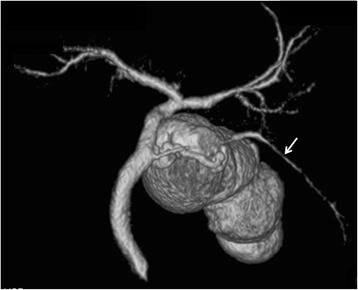
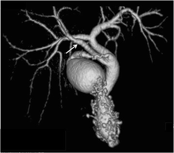
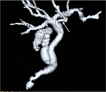
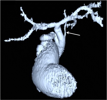
Methods: Five hundred sixty nine patients who underwent preoperative DIC-CT and laparoscopic cholecystectomy were reviewed. Accessory hepatic ducts were classified as follows: type I (accessory hepatic ducts that merged with the common hepatic duct between the confluence of the right and left hepatic ducts and the cystic duct confluence), type II (those that merged with the common hepatic duct at the same site as the cystic duct), type III (those that merged with the common bile duct distal to the cystic duct confluence), type IV (the cystic duct merged with the accessory hepatic duct), and type V (accessory hepatic ducts that merged with the common hepatic or bile duct on the left side).

Results: Accessory hepatic ducts were observed in 50 patients. Type I, II, III, IV, and V accessory hepatic ducts were detected in 32, 3, 1, 11, and 3 patients, respectively. Based on their drainage areas, the accessory hepatic ducts were also classified as follows: a posterior branch in 22 patients, an anterior branch in 9 patients, a combination of posterior and anterior branches in 16 patients, a left-sided branch in 2 patients, and a caudate branch in 1 patient. None of the patients with accessory hepatic ducts suffered bile duct injuries.

Conclusion: There are a number of variants of the accessory hepatic duct. DIC-CT is useful to detect the accessory hepatic duct.

Keywords: Accessory hepatic duct; Bile duct injury; Drip infusion cholangiography with CT; Laparoscopic cholecystectomy.


Figures

Fig. 1
Fig. 1
Classification of accessory hepatic ducts. The arrow indicates the accessory hepatic duct, and the arrow head shows the cystic duct
Fig. 2
Fig. 2
Three-dimensional DIC-CT (right ventral view) showing a type I accessory hepatic duct. The accessory hepatic duct (arrow) drained both the dorsal area of the right anterior section (white arrow head) and the right posterior section (black arrow head) and was classified into the combined posterior and anterior branch type
Fig. 3
Fig. 3
Three-dimensional DIC-CT image (dorsal view) showing a type II accessory hepatic duct. The accessory hepatic duct drained segment 6 (arrow) and was classified as belonging to the posterior branch type
Fig. 4
Fig. 4
Three-dimensional DIC-CT image (ventral view) showing a type III accessory hepatic duct. The accessory hepatic duct drained the right anterior section (arrow) and was classified as belonging to the anterior branch type
Fig. 5
Fig. 5
Three-dimensional DIC-CT image (ventral view) showing a type IV accessory hepatic duct. The accessory hepatic duct drained the right posterior section (arrow) and was classified as belonging to the posterior branch type
Fig. 6
Fig. 6
Three-dimensional DIC-CT image (ventral view) showing the type V accessory hepatic duct. The accessory hepatic duct drained segment 4 (arrow) and was classified into the left-sided branch type
Fig. 7
Fig. 7
Three-dimensional DIC-CT image (ventral view) showing a type V accessory hepatic duct. The accessory hepatic duct drained the Spiegel lobe (arrow) and was classified into the caudate branch type

Similar articles

Cited by

References

    1. Uchiyama K, Tani M, Kawai M, Ueno T, Hama T, Yamaue H. Preoperative evaluation of the extrahepatic bile duct structure for laparoscopic cholecystectomy. Surg Endosc. 2006;20:1119–1123. doi: 10.1007/s00464-005-0689-1. - DOI - PubMed
    1. Nijssen MA, Schreinemakers JM, Meyer Z, van der Schelling GP, Crolla RM, Rijken AM. Complication after laparoscopic cholecystectomy: a video evaluation study of whether the critical view of safety was reached. World J Surg. 2015;39:1798–1803. doi: 10.1007/s00268-015-2993-9. - DOI - PubMed
    1. Hugh TB. New strategies to prevent laparoscopic bile duct injury: surgeons can learn from pilots. Surgery. 2002;132:826–835. doi: 10.1067/msy.2002.127681. - DOI - PubMed
    1. Noji T, Nakamura F, Nakamura T, Kato K, Suzuki O, Ambo Y, et al. ENBD tube placement prior to laparoscopic cholecystectomy may reduce the rate of complications in cases with predictably complicating biliary anomalies. J Gastroenterol. 2011;46:73–77. doi: 10.1007/s00535-010-0281-x. - DOI - PubMed
    1. Ochiai T, Yamazaki S, Ohta K, Takahashi M, Iwai T, Irie T, et al. Is drip infusion cholecystocholangiography (DIC) an acceptable modality at cholecystectomy for cholecystolithiasis, considering the frequency of bile duct maljunction and intraoperative bile duct injury? J Hepato-Biliary-Pancreat Surg. 2004;11:135–139. doi: 10.1007/s00534-003-0873-4. - DOI - PubMed

MeSH terms