. 2017 May 8;18(1):87.
doi: 10.1186/s12931-017-0571-4.

Selective gene expression analysis of the neuroepithelial body microenvironment in postnatal lungs with special interest for potential stem cell characteristics

Affiliations

Selective gene expression analysis of the neuroepithelial body microenvironment in postnatal lungs with special interest for potential stem cell characteristics

Line Verckist et al. Respir Res. .

Abstract

Background: The pulmonary neuroepithelial body (NEB) microenvironment (ME) consists of innervated cell clusters that occur sparsely distributed in the airway epithelium, an organization that has so far hampered reliable selective gene expression analysis. Although the NEB ME has been suggested to be important for airway epithelial repair after ablation, little is known about their potential stem cell characteristics in healthy postnatal lungs. Here we report on a large-scale selective gene expression analysis of the NEB ME.

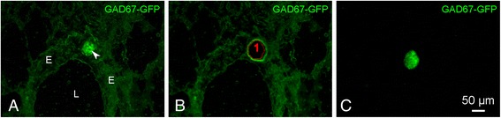
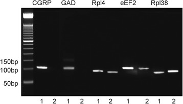
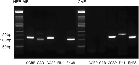
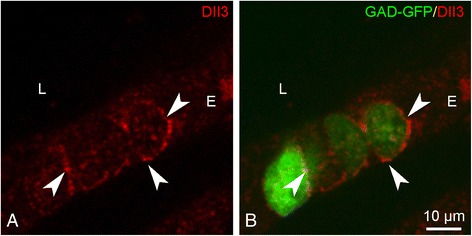
Methods: A GAD67-GFP mouse model was used that harbors GFP-fluorescent NEBs, allowing quick selection and pooling by laser microdissection (LMD) without further treatment. A panel of stem cell-related PCR arrays was used to selectively compare mRNA expression in the NEB ME to control airway epithelium (CAE). For genes that showed a higher expression in the NEB ME, a ranking was made based on the relative expression level. Single qPCR and immunohistochemistry were used to validate and quantify the PCR array data.

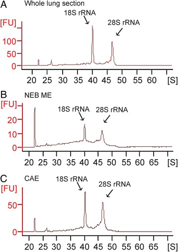
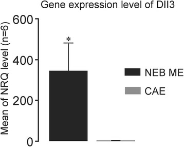
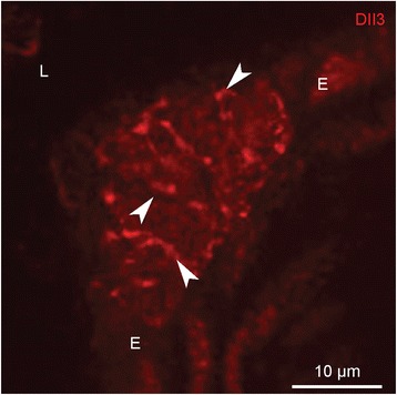
Results: Careful optimization of all protocols appeared to be essential to finally obtain high-quality RNA from pooled LMD samples of NEB ME. About 30% of the more than 600 analyzed genes showed an at least two-fold higher expression compared to CAE. The gene that showed the highest relative expression in the NEB ME, Delta-like ligand 3 (Dll3), was investigated in more detail. Selective Dll3 gene expression in the NEB ME could be quantified via single qPCR experiments, and Dll3 protein expression could be localized specifically to NEB cell surface membranes.

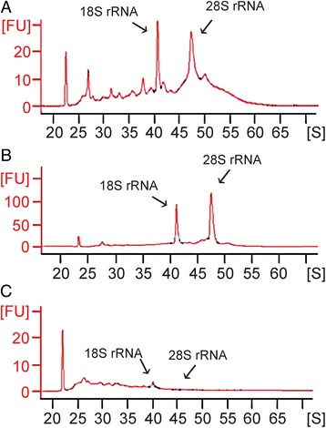
Conclusions: This study emphasized the importance of good protocols and RNA quality controls because of the, often neglected, fast RNA degradation in postnatal lung samples. It was shown that sufficient amounts of high-quality RNA for reliable complex gene expression analysis can be obtained from pooled LMD-collected NEB ME samples of postnatal lungs. Dll3 expression, which has also been reported to be important in high-grade pulmonary tumor-initiating cells, was used as a proof-of-concept to confirm that the described methodology represents a promising tool for further unraveling the molecular basis of NEB ME physiology in general, and its postnatal stem cell capacities in particular.

Keywords: Airway epithelium; Delta-like ligand 3; Laser microdissection; Neuroepithelial body microenvironment; PCR array; Pulmonary neuroendocrine cells; Stem cell niche.


Figures

Fig. 1
Fig. 1
Example of the isolation of a pulmonary NEB from a GAD67-GFP mouse lung cryosection using LMD (see Methods). a GFP-fluorescent NEB (arrowhead) located in the epithelium of an intrapulmonary airway. b Image taken after LMD. The area circled by the red line shows the region of interest that was selected to be cut by the laser. c Isolated GFP-fluorescent NEB, captured in the cap of an Eppendorf tube and ready for consecutive pooling and RNA isolation. Note that even after very mild fixation, to optimally preserve RNA quality, and without cover glass, NEBs appear to be unambiguously detectable in the LMD microscope (Leica LMD7000; 20x objective). L: airway lumen, E: airway epithelium
Fig. 2
Fig. 2
Electropherograms demonstrating the 18S and 28S rRNA peaks, corresponding to the level of intact RNA in each sample, are used for total RNA quality analysis of random LMD-collected and pooled small samples from cryostat sections of brain (PD21; a), embryonic (ED14; b) and postnatal lung (PD21, c). In the brain (RNA Quality Indicator, RIN = 7.9) and embryonic mouse lung (RIN = 8.9), high quality intact RNA can be detected, while in the identically processed postnatal mouse lung tissue a large part of the RNA appears to be degraded (RIN = 3.2)
Fig. 3
Fig. 3
Electropherograms of a postnatal whole lung LMD section (a), pooled NEB ME (b) and CAE (c) samples that were obtained after optimized tissue processing, LMD and RNA isolation protocols. For all samples, the concentration of RNA was calculated by determining the area under the entire RNA electropherogram
Fig. 4
Fig. 4
Non-amplified RNA samples. Detection of CGRP, GAD and reference genes’ expression in NEB ME (lane 1) and in a whole lung section (lane 2) of a GAD67-GFP mouse (PD21). The amplicon length of CGRP is 105 bp and that of GAD is 102 bp, both clearly visible as a distinct band on the gel electrophoresis image. For the whole lung sample, there is no visible band that can account for the CGRP or GAD amplicon, while there are positive bands for the NEB RNA sample. The reference genes are expressed in every sample
Fig. 5
Fig. 5
Gene expression of CGRP, GAD, CCSP, Flt-1 and Rpl38 in amplified samples of the NEB ME and CAE. CGRP and GAD are selective markers for neuroendocrine cells and gene expression can be seen in the NEB ME only. CCSP is a marker for both CLCs and Clara cells, and gene expression can be seen in NEB ME and CAE samples. Flt-1 is a selective marker for ciliated cells, which are present in the CAE sample only. Rpl38 is shown as reference gene and is strongly expressed in the NEB ME and in CAE
Fig. 6
Fig. 6
Results of qPCR experiments for the expression of Dll3. The mean expression in LMD samples of the NEB ME of five different mice is compared with matched CAE samples. The NRQ shows Dll3 mRNA levels in the NEB ME, as compared to the levels in the CAE, and normalized to expression of the reference genes Rpl38, eEF2 and Rpl4 in the same five samples. NRQ: Normalized Relative Quantification, n = 5. Differences are considered significant: p < 0,05 (*), using Mann-Whitney U test
Fig. 7
Fig. 7
Single immunostaining for Dll3 (red Cy3 fluorescence) in a cryosection of the lungs of a wildtype mouse (PD21). Dll3 shows a spot-like staining (arrowheads) at distinct locations in the airway epithelium, covering the surface membrane of groups of intraepithelial cells. L: airway lumen, E: airway epithelium
Fig. 8
Fig. 8
Dll3 immunostaining (red Cy3 fluorescence; a) on a lung cryosection of a GAD67-GFP mouse that shows GFP-expressing (green GFP fluorescence; b) NEB cells. Typically a spot-like Dll3 staining of the surface membrane of NEB cells can be observed (arrowheads). L: airway lumen, E: airway epithelium

Similar articles

Cited by

References

    1. Adriaensen D, Scheuermann DW. Neuroendocrine cells and nerves of the lung. Anat Rec. 1993;236:70–86. doi: 10.1002/ar.1092360111. - DOI - PubMed
    1. Stahlman MT, Gray ME. Ontogeny of neuroendocrine cells in human-fetal lung 1. An electron-microscopic study. Lab Invest. 1984;51:449–63. - PubMed
    1. Haller CJ. A scanning and transmission electron-microscopic study of the development of the surface-structure of neuroepithelial bodies in the mouse lung. Micron. 1994;25:527–38. doi: 10.1016/0968-4328(94)90015-9. - DOI - PubMed
    1. Reynolds SD, Giangreco A, Power JHT, Stripp BR. Neuroepithelial bodies of pulmonary airways serve as a reservoir of progenitor cells capable of epithelial regeneration. Am J Pathol. 2000;156:269–78. doi: 10.1016/S0002-9440(10)64727-X. - DOI - PMC - PubMed
    1. Linnoila RI. Functional facets of the pulmonary neuroendocrine system. Lab Invest. 2006;86:425–44. doi: 10.1038/labinvest.3700412. - DOI - PubMed

Publication types

Substances