. 2017 May 8;17(1):319.
doi: 10.1186/s12885-017-3246-0.

Effect of estrogen receptor β agonists on proliferation and gene expression of ovarian cancer cells

Affiliations

Effect of estrogen receptor β agonists on proliferation and gene expression of ovarian cancer cells

Susanne Schüler-Toprak et al. BMC Cancer. .

Abstract

Background: Estrogen receptor (ER) β has been suggested to affect ovarian carcinogenesis. We examined the effects of four ERβ agonists on proliferation and gene expression of two ovarian cancer cell lines.

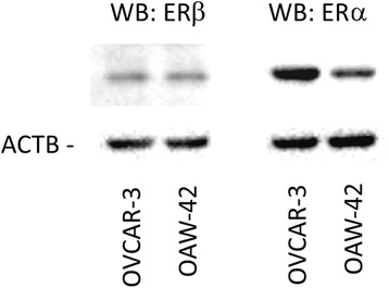
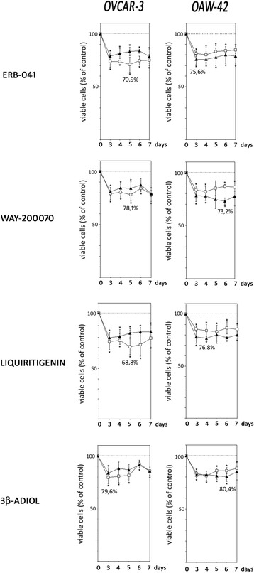
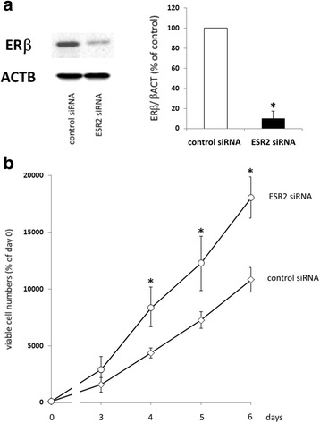
Methods: OVCAR-3 and OAW-42 ovarian cancer cells were treated with the ERβ agonists ERB-041, WAY200070, Liquiritigenin and 3β-Adiol and cell growth was measured by means of the Cell Titer Blue Assay (Promega). ERβ expression was knocked down by transfection with specific siRNA. Additionally, transcriptome analyses were performed by means of Affymetrix GeneChip arrays. To confirm the results of DNA microarray analysis, Western blot experiments were performed.

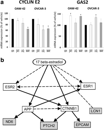
Results: All ERβ agonists tested significantly decreased proliferation of OVCAR-3 and OAW-42 cells at a concentration of 10 nM. Maximum antiproliferative effects were induced by flavonoid Liquiritigenin, which inhibited growth of OVCAR-3 cells by 31.2% after 5 days of treatment, and ERB-041 suppressing proliferation of the same cell line by 29.1%. In OAW-42 cells, maximum effects were observed after treatment with the ERβ agonist WAY200070, inhibiting cell growth by 26.8%, whereas ERB-041 decreased proliferation by 24.4%. In turn, knockdown of ERβ with specific siRNA increased cell growth of OAW-42 cells about 1.9-fold. Transcriptome analyses revealed a set of genes regulated by ERβ agonists including ND6, LCN1 and PTCH2, providing possible molecular mechanisms underlying the observed antiproliferative effects.

Conclusion: In conclusion, the observed growth-inhibitory effects of all ERβ agonists on ovarian cancer cell lines in vitro encourage further studies to test their possible use in the clinical setting.

Keywords: Estrogen receptor beta; Estrogen receptor beta agonists; Ovarian cancer.


Figures

Fig. 1
Fig. 1
Expression of ERβ and ERα in OVCAR-3 and OAW-42 ovarian cancer cells. Expression of the indicated receptors was examined by means of Western blot analysis. Levels of β-Actin (AKTB) were determined as internal control. Aliquots containing 10 μg of protein isolated from both cell lines were resolved by 10% (w/v) SDS–polyacrylamide gel electrophoresis, followed by electrotransfer to a PVDF hybond membrane (Amersham, UK)
Fig. 2
Fig. 2
Effects of ERβ-agonists on growth of OVCAR-3 and OAW-42 ovarian cancer cells. OVCAR-3 and OAW-42 cells cultured in medium containing 10% FCS (open squares) or defined serum replacement SR2 (filled triangles) were treated with 10 nM of ERB-041, WAY-200070, Liquiritigenin or 3β-Adiol as indicated for up to 7 days and relative numbers of viable cells were determined by means of the fluorimetric CellTiter-Blue® Assay (Promega). Data are expressed in percent of the vehicle controls (n = 4; * P < 0.05 vs. control; ** P < 0.01 vs. control)
Fig. 3
Fig. 3
Effect of an ERβ knockdown on proliferation of OAW-42 cells. a: ERβ expression in OAW-42 ovarian cancer cells after transfection with ERβ siRNA compared to controls. 72 h after transfection, total protein was isolated and knockdown was examined on the protein level by means of Western blot analysis as described in the methods section. ERβ expression levels after transfection with a mix of ESR2 siRNAs (10 nM each) were compared to levels in cells transfected with negative control siRNA (n = 4). *p < 0.01 vs. control-transfected cells. b: Proliferation of OAW-42 cells with reduced levels of ERβ. Cells were transfected with ESR2-specific siRNA or negative control siRNA and seeded into 96-well plates (1000 cells/well) in medium containing 10% FCS the next day. 0, 3, 4, 5, and 6 days after transfection, relative numbers of viable cells were determined by means of the fluorimetric CellTiter-Blue® Assay (Promega). From one vial of transfected cells, 72 h after transfection total RNA and protein was isolated in parallel to confirm knockdown of ESR2 expression. Data are expressed in percent of day 0 (n = 4). *p < 0.01 vs. control-transfected cells
Fig. 4
Fig. 4
Western blot analysis demonstrating down-regulated protein expression of the indicated genes after treatment with the ERβ agonists ERβ-041, WAY200070 and Liquiritigenin. 72 h after stimulation with 10 nM of the agonists, total protein was isolated and subjected to Western blot analysis. Analyses were performed using specific antibodies against the gene products of LCN1, ND6 and PTCH2 and additionally ACTB as a loading control. Shown are representative results and the densitometrical mean values in relation to ACTB (n = 3). *p < 0.01 vs. vehicle
Fig. 5
Fig. 5
Effect of ERβ agonists on gene expression (Affymetrix GeneChip analysis). a Regulation of growth-associated genes cyclin E2 and growth arrest specific 2 (GAS2) after treatment with the agonists ERB-041 (β41), Liquiritigenin (LIQ), WAY200070 (WAY) or the vehicle control (con) for 48 h (10 nM) *p < 0.05 bs. Vehicle. b Network connecting ESR2 with the genes LCN1, EPCAM, PTCH2 and ND6 being downregulated by ERβ agonists in this study. Broken lines: direct binding. Solid lines: affecting expression. Prediction by IPA Software (Ingenuity Pathway Analysis, Ingenuity Systems, Stanford, USA) [–61]

Similar articles

Cited by

References

    1. Siegel RL, Miller KD, Jemal A. Cancer statistics, 2015. CA Cancer J Clin. 2015;65(1):5–29. doi: 10.3322/caac.21254. - DOI - PubMed
    1. Aikhionbare FO, Mehrabi S, Kumaresan K, Zavareh M, Olatinwo M, Odunsi K, Partridge E. Mitochondrial DNA sequence variants in epithelial ovarian tumor subtypes and stages. J Carcinog. 2007;6:1. doi: 10.1186/1477-3163-6-1. - DOI - PMC - PubMed
    1. Docquier A, Garcia A, Savatier J, Boulahtouf A, Bonnet S, Bellet V, Busson M, Margeat E, Jalaguier S, Royer C, et al. Negative regulation of estrogen signaling by ERbeta and RIP140 in ovarian cancer cells. Mol Endocrinol. 2013;27(9):1429–1441. doi: 10.1210/me.2012-1351. - DOI - PMC - PubMed
    1. Greenlee RT, Murray T, Bolden S, Wingo PA. Cancer statistics, 2000. CA Cancer J Clin. 2000;50(1):7–33. doi: 10.3322/canjclin.50.1.7. - DOI - PubMed
    1. Havrilesky LJ, McMahon CP, Lobenhofer EK, Whitaker R, Marks JR, Berchuck A. Relationship between expression of coactivators and corepressors of hormone receptors and resistance of ovarian cancers to growth regulation by steroid hormones. J Soc Gynecol Investig. 2001;8(2):104–113. doi: 10.1016/S1071-5576(01)00093-4. - DOI - PubMed

MeSH terms