. 2017 May 8;19(1):87.
doi: 10.1186/s13075-017-1300-6.

Disturbed microcirculation in the hands of patients with systemic sclerosis detected by fluorescence optical imaging: a pilot study

Affiliations

Disturbed microcirculation in the hands of patients with systemic sclerosis detected by fluorescence optical imaging: a pilot study

Stefanie Friedrich et al. Arthritis Res Ther. .

Abstract

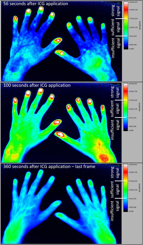
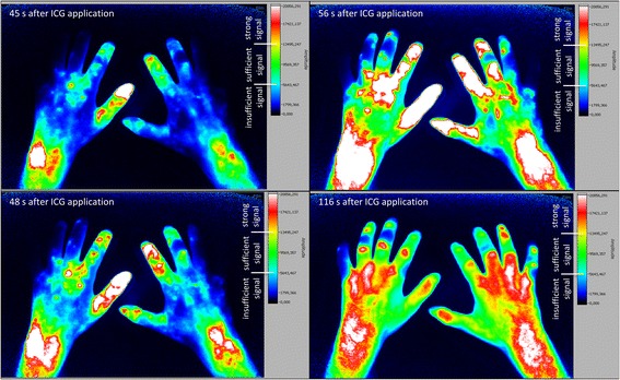
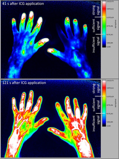
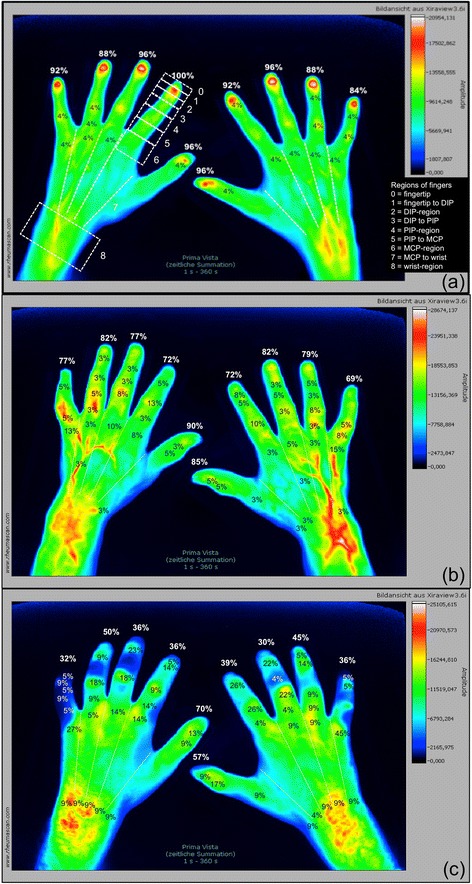
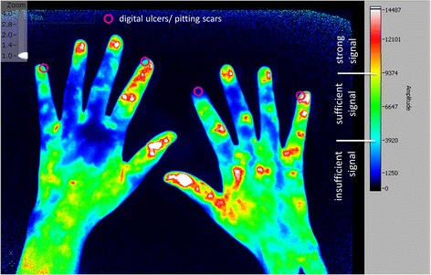
Background: Utilising fluorescence optical imaging (FOI), the distribution of an intravenously applied colouring agent indocyanine green (ICG) can be analysed with the potential to identify malperfusion by little to no tissue enhancement. Systemic sclerosis (SSc) is characterised by the presence of digital ulcers reflecting progressive vasculopathy. The objective was to investigate the potential of FOI in the detection of disturbed microcirculation in the hands and fingers of patients with SSc and to link FOI findings to clinical signs of ischemia such as digital ulcers and pitting scars.

Methods: In this cross-sectional study, 63 patients with SSc and 26 healthy subjects were examined. FOI was performed in all 89 individuals and compared to clinical data and capillaroscopic findings assembled for the SSc cohort.

Results: Healthy subjects showed initial ICG signals in their fingertips in 93.6%, SSc patients in 78.5% (limited SSc) and 43.2% (diffuse SSc). Moreover, in SSc patients, FOI findings were significantly associated with a late capillaroscopic pattern, disseminated SSc features, a diffuse SSc subtype, and the presence of digital ulcers or pitting scars. Intra- and inter-reader reliability for FOI amounted to κ = 0.786 and κ = 0.834, respectively.

Conclusions: FOI is able to detect areas of reduced microcirculation in patients with SSc with high association to capillaroscopic findings. The results pave the way for future FOI investigations into its role in the prediction of complications due to an impaired acral perfusion.

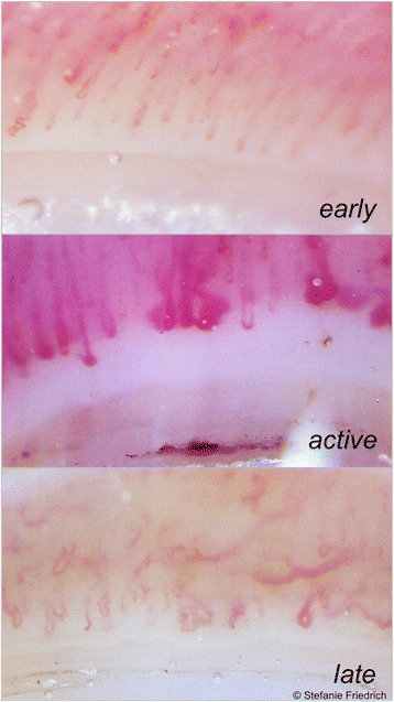
Keywords: Digital ulcers; Disturbed microcirculation; Fluorescence optical imaging; Nailfold capillaroscopy; Raynaud’s phenomenon; Systemic sclerosis.


Figures

Fig. 1
Fig. 1
Fluorescence optical imaging (FOI) staining in a healthy subject. Upper image: initial enhancement (IE), defined as the first strong signal of indocyanine green (ICG), in digit II of the right hand; IE in the left hand has already taken place. Middle image: area of maximum distal distribution (MDD) is the fingertip (region 0) in every finger as this is the most distal region with a sufficient ICG signal. Lower image: last image (no. 360) of the examination; most of the ICG has already disappeared
Fig. 2
Fig. 2
Fluorescence optical imaging (FOI) staining in a patient with diffuse cutaneous systemic sclerosis. Upper left image: strong initial enhancement (IE) in digit II of the right hand just below the fingertip (region 1). Lower left image: IE in the digit III of the left hand near the metacarpophalangeal (MCP) joint (region 6). Upper right image: maximum distal distribution (MDD) in the fingertip (region 0) of digit II of the right hand. Lower right image: MDD in digit V of both hands with the most distal sufficient signal in region 5 of the left digit V and region 1 of the right hand’s digit V. ICG indocyanine green
Fig. 3
Fig. 3
Fluorescence optical imaging (FOI) staining in a patient with lcSSc, without clinically apparent Raynaud’s episode. Upper image: strong initial enhancement (IE) in digits I, III, IV and V of the right hand; the index finger has not yet accumulated indocyanine green (ICG). Lower image: maximum distal distribution (MDD) in the fingertip of the right hand’s index finger
Fig. 4
Fig. 4
Regions of initial enhancement in healthy subjects and systemic sclerosis patients. PrimaVistaMode (summation image) of (a) a healthy subject and exemplary depiction of the nine regions in digit II, left hand with finger-wise percentage of initial enhancement of indocyanine green (ICG) of the healthy cohort: proper distribution of the colouring agent with 84–100% (mean: 93.1%) of first ICG signals in region 0 (fingertip). PrimaVistaMode of a patient with (b) limited (cutaneous) systemic sclerosis (lcSSc) and (c) diffuse cutaneous systemic sclerosis (dcSSc) and depiction of the finger-wise percentage of strong initial ICG enhancement per region (0–8). LcSSc cohort with 69–90% (mean: 78.5%) and dcSSc cohort with 30–70% (mean: 43.2%) of initial ICG signals in region 0. DIP distal interphalangeal joint, MCP metacarpophalangeal joint, PIP proximal interphalangeal joint
Fig. 5
Fig. 5
Typical nailfold capillaroscopy patterns in systemic sclerosis patients. Exemplary images of the three patterns of nailfold capillaroscopy as defined by Cutolo et al. [29]: the early pattern (upper image) is characterised by a normal capillary density and distribution with few ectasias and haemorrhages. The active pattern (middle image) is defined by giant capillaries, many haemorrhages and mild capillary loss and disorganisation. Irregular capillary width, avascular areas due to decreased capillary density as well as ramified or bushy capillaries, but less giant capillaries and haemorrhages constitute the late pattern (lower image)
Fig. 6
Fig. 6
Location of digital ulcers and pitting scars in patients with pathologic FOI. Exemplary depiction of the location of current digital ulcer and/or pitting scars (DU/PS) projected onto a fluorescence optical imaging frame 62 seconds after application of indocyanine green (ICG). The initial enhancement (IE) has already taken place in all fingers with the first strong signal proximal to region 0 in digit II of the right hand

Similar articles

Cited by

References

    1. Werner SG, Langer HE, Schott P, et al. Indocyanine green-enhanced fluorescence optical imaging in patients with early and very early arthritis: a comparative study with magnetic resonance imaging. Arthritis Rheum. 2013;65(12):3036–44. doi: 10.1002/art.38175. - DOI - PubMed
    1. Glimm AM, Werner SG, Burmester GR, et al. Analysis of distribution and severity of inflammation in patients with osteoarthitis compared to rheumatoid arthritis by ICG-enhanced fluorescence optical imaging and musculoskeletal ultrasound: a pilot study. Ann Rheum Dis. 2016;75(3):566–70. doi:10.1136/annrheumdis-2015-207345. - PMC - PubMed
    1. Fischer T, Ebert B, Voigt J, et al. Detection of rheumatoid arthritis using non-specific contrast enhanced fluorescence imaging. Acad Radiol. 2010;17(3):375–81. doi: 10.1016/j.acra.2009.09.016. - DOI - PubMed
    1. Mothes H, Dönicke T, Friedel R, et al. Indocyanine-green fluorescence video angiography used clinically to evaluate tissue perfusion in microsurgery. J Trauma. 2004;57(5):1018–24. doi: 10.1097/01.TA.0000123041.47008.70. - DOI - PubMed
    1. Benya R, Quintana J, Brundage B. Adverse reactions to indocyanine green: a case report and a review of the literature. Cathet Cardiovasc Diagn. 1989;17:231–3. doi: 10.1002/ccd.1810170410. - DOI - PubMed

Publication types