. 2017 May 8;8(1):93.
doi: 10.1186/s13287-017-0538-x.

Critical attributes of human early mesenchymal stromal cell-laden microcarrier constructs for improved chondrogenic differentiation

Affiliations

Critical attributes of human early mesenchymal stromal cell-laden microcarrier constructs for improved chondrogenic differentiation

Youshan Melissa Lin et al. Stem Cell Res Ther. .

Abstract

Background: Microcarrier cultures which are useful for producing large cell numbers can act as scaffolds to create stem cell-laden microcarrier constructs for cartilage tissue engineering. However, the critical attributes required to achieve efficient chondrogenic differentiation for such constructs are unknown. Therefore, this study aims to elucidate these parameters and determine whether cell attachment to microcarriers throughout differentiation improves chondrogenic outcomes across multiple microcarrier types.

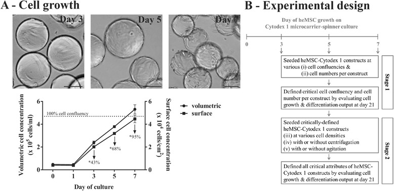
Methods: A screen was performed to evaluate whether 1) cell confluency, 2) cell numbers, 3) cell density, 4) centrifugation, or 5) agitation are crucial in driving effective chondrogenic differentiation of human early mesenchymal stromal cell (heMSC)-laden Cytodex 1 microcarrier (heMSC-Cytodex 1) constructs.

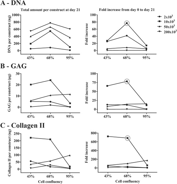
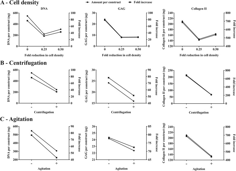
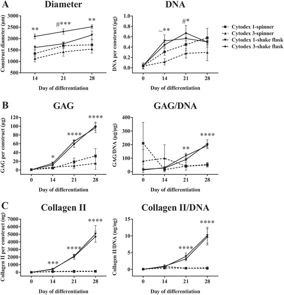
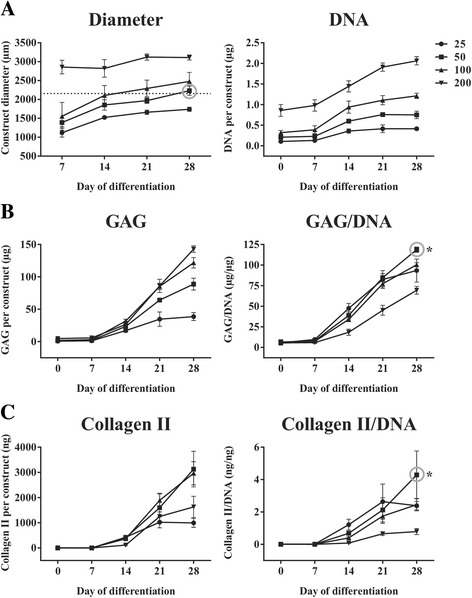
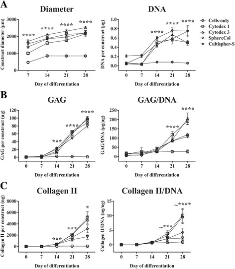
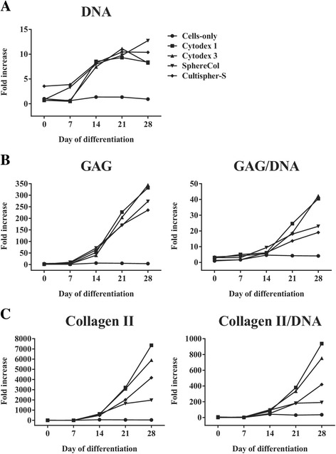
Results: Firstly, we found that seeding 10 × 103 cells at 70% cell confluency with 300 microcarriers per construct resulted in substantial increase in cell growth (76.8-fold increase in DNA) and chondrogenic protein generation (78.3- and 686-fold increase in GAG and Collagen II, respectively). Reducing cell density by adding empty microcarriers at seeding and indirectly compacting constructs by applying centrifugation at seeding or agitation throughout differentiation caused reduced cell growth and chondrogenic differentiation. Secondly, we showed that cell attachment to microcarriers throughout differentiation improves cell growth and chondrogenic outcomes since critically defined heMSC-Cytodex 1 constructs developed larger diameters (2.6-fold), and produced more DNA (13.8-fold), GAG (11.0-fold), and Collagen II (6.6-fold) than their equivalent cell-only counterparts. Thirdly, heMSC-Cytodex 1/3 constructs generated with cell-laden microcarriers from 1-day attachment in shake flask cultures were more efficient than those from 5-day expansion in spinner cultures in promoting cell growth and chondrogenic output per construct and per cell. Lastly, we demonstrate that these critically defined parameters can be applied across multiple microcarrier types, such as Cytodex 3, SphereCol and Cultispher-S, achieving similar trends in enhancing cell growth and chondrogenic differentiation.

Conclusions: This is the first study that has identified a set of critical attributes that enables efficient chondrogenic differentiation of heMSC-microcarrier constructs across multiple microcarrier types. It is also the first to demonstrate that cell attachment to microcarriers throughout differentiation improves cell growth and chondrogenic outcomes across different microcarrier types, including biodegradable gelatin-based microcarriers, making heMSC-microcarrier constructs applicable for use in allogeneic cartilage cell therapy.

Keywords: Cartilage; Cell therapy; Chondrogenic differentiation; Mesenchymal stromal cells; Microcarrier.


Figures

Fig. 1
Fig. 1
Evaluation of critical parameters required to achieve efficient chondrogenic differentiation of heMSC-Cytodex 1 microcarrier constructs. a Brightfield images (scale bar = 100 μm) and kinetics of heMSC growth on Cytodex 1 microcarriers in agitated spinner culture. Numbers indicate the cell confluency (dotted line represents 100% cell confluency of 4.7 × 104 cells/cm2 as calculated from monolayer cultures). *Cell-laden microcarriers taken from spinner culture at the indicated time point were used to seed heMSC-Cytodex 1 constructs. b Schematic of experimental design. Stage 1: heMSC attached to Cytodex 1 microcarriers were seeded as chondrogenic heMSC-microcarrier constructs at either day 3 (early-log phase with 43% cell confluency), day 5 (mid-log phase with 68% cell confluency), or day 7 (late-log phase with 95% cell confluency), using different cell numbers per construct. Stage 2: heMSC-microcarrier constructs generated under critically defined conditions as identified at Stage 1 were evaluated for the effect of cell density (addition of empty microcarriers at seeding) or the effect of compaction (centrifugation at seeding or agitation throughout differentiation)
Fig. 2
Fig. 2
Seeding 10 × 103 cells at 68% cell confluency per heMSC-Cytodex 1 construct (grey circle) resulted in efficient cell growth and chondrogenic differentiation by 21 days of differentiation. a DNA, b GAG, and c Collagen II content per construct by day 21 of differentiation as well as respective fold-increases from day 0 to day 21 of differentiation
Fig. 3
Fig. 3
Reduction in cell density by adding empty microcarriers at seeding and construct compaction by applying centrifugation at seeding or continuous agitation throughout differentiation had a negative impact on cell growth and chondrogenic output. a DNA, b GAG, and c Collagen II content per construct at day 21 of differentiation and relevant fold-increases from day 0 to day 21 of differentiation
Fig. 4
Fig. 4
heMSC-Cytodex 1 constructs developed larger pellet diameters, increased cellular proliferation, and, most importantly, improved total chondrogenic output in terms of proteoglycan and Collagen II content as compared to their equivalent cell-only counterparts. a Kinetics of cell growth and chondrogenic differentiation. heMSC-Cytodex 1 constructs and cell-only pellets were seeded with 10 × 103 heMSC at 70% cell confluency. Kinetics of construct/pellet diameter, DNA, GAG, and Collagen II production were monitored during 28 days of differentiation. All p values refer to statistical significance obtained by comparing heMSC-Cytodex 1 constructs to that of cell-only counterparts at the indicated time points. p values: n.s. = p > 0.05, *p < 0.05, **p < 0.01, ***p < 0.001. and ****p < 0.0001. All numbers shown indicate the fold-changes of heMSC-Cytodex 1 constructs over that of cell-only pellets at the indicated time points. b Histological H&E, Safranin O, Alcian Blue, and Collagen II staining of heMSC-Cytodex 1 constructs and cell-only pellets at day 28 of differentiation. Arrows indicate areas in heMSC-Cytodex 1 constructs with more intense staining compared to that of cell-only pellets. The space occupied by the microcarrier is indicated as “mc”. Scale bar = 500 μm
Fig. 5
Fig. 5
heMSC-Cytodex 1/3 constructs created via agitated shake flask platform were more efficient in promoting cell growth as well as chondrogenic output per construct and per cell as compared to those derived from agitated spinner platform. a Construct diameter and DNA content per construct. b GAG content per construct and GAG/DNA ratio. c Collagen II content per construct and Collagen II/DNA ratio. p values: *p < 0.05, **p < 0.01, ***p < 0.001, and ****p < 0.0001. All p values refer to the statistical significance of heMSC-Cytodex 1/3 constructs from agitated shake flask platform compared to those derived from spinner platform at the indicated time points. #All comparisons except between Cytodex 1-shake flask and Cytodex 1-spinner. ~All comparisons except between Cytodex 3-shake flask and Cytodex 1-spinner
Fig. 6
Fig. 6
heMSC-Cultispher-S constructs seeded with 50 microcarriers per construct with 70% cell confluency induced effective cell growth and chondrogenic differentiation. a Construct diameter and DNA content per construct. Dotted line indicates construct diameter attained by optimally defined heMSC-Cytodex 1 constructs after 28 days of differentiation. This diameter is obtained by heMSC-Cultispher-S constructs seeded with 50 microcarriers per construct (grey circle). b, c Glycosaminogycan (GAG) and Collagen II content per construct as well as GAG/DNA and Collagen II/DNA ratios. heMSC-Cultispher-S constructs seeded with 50 microcarriers (grey circle) significantly induced the highest GAG/DNA and Collagen II/DNA ratios at day 28 of differentiation when compared to constructs with different microcarrier numbers. p values: *p < 0.05
Fig. 7
Fig. 7
heMSC-microcarrier constructs of different microcarrier types increased cellular proliferation and improved chondrogenic output per construct and per cell when compared to their equivalent cell-only counterparts. a Construct diameter and DNA content per construct. b Glycosaminogycan (GAG) content per construct and GAG/DNA ratio. c Collagen II content per construct and Collagen II/DNA ratio. p values, *p < 0.05, ***p < 0.001, and ****p < 0.0001. All p values refer to the statistical significance of all heMSC-microcarrier constructs across distinct microcarrier types over that of cell-only counterparts at the indicated time points. ~All heMSC-microcarrier pellets except that of SphereCol and Cultispher-S
Fig. 8
Fig. 8
heMSC-microcarrier constructs of different microcarrier types increased cellular proliferation and improved chondrogenic output per construct and per cell when compared to their equivalent cell-only counterparts. Fold-increases of a DNA content per construct, b Glycosaminogycan (GAG) content per construct and GAG/DNA ratio, and c Collagen II content per construct and Collagen II/DNA ratio across different microcarrier types over the cell-only value at day 0 of differentiation

Similar articles

Cited by

References

    1. Sophia Fox AJ, Bedi A, Rodeo SA. The basic science of articular cartilage: structure, composition, and function. Sports Health. 2009;1:461–8. doi: 10.1177/1941738109350438. - DOI - PMC - PubMed
    1. Jasin HE. Structure and function of the articular cartilage surface. Scand J Rheumatol Suppl. 1995;101:51–5. doi: 10.3109/03009749509100900. - DOI - PubMed
    1. Buckwalter JA, Mankin HJ. Articular cartilage: tissue design and chondrocyte-matrix interactions. Instr Course Lect. 1998;47:477–86. - PubMed
    1. Bhosale AM, Richardson JB. Articular cartilage: structure, injuries and review of management. Br Med Bull. 2008;87:77–95. doi: 10.1093/bmb/ldn025. - DOI - PubMed
    1. Buckwalter JAMH. Articular cartilage, part 1: tissue design and chondrocyte-matrix interaction. J Bone Joint Surg Am. 1997;79:600–11. doi: 10.2106/00004623-199704000-00021. - DOI