

Langerhans cell histiocytosis of the maxillae in a child treated only with chemotherapy: a case report
- PMID: 28482919
- PMCID: PMC5422897
- DOI: 10.1186/s13256-017-1286-3
Langerhans cell histiocytosis of the maxillae in a child treated only with chemotherapy: a case report
Abstract
Background: Langerhans cell histiocytosis is a sporadic disease caused by an uncontrolled pathogenic clonal proliferation of dendritic cells that have Langerhans cell characteristics. New treatment protocols provided by the HISTSOC-LCH-III (NCT00276757) trial show an improvement in the survival of children with langerhans cell histiocytosis.
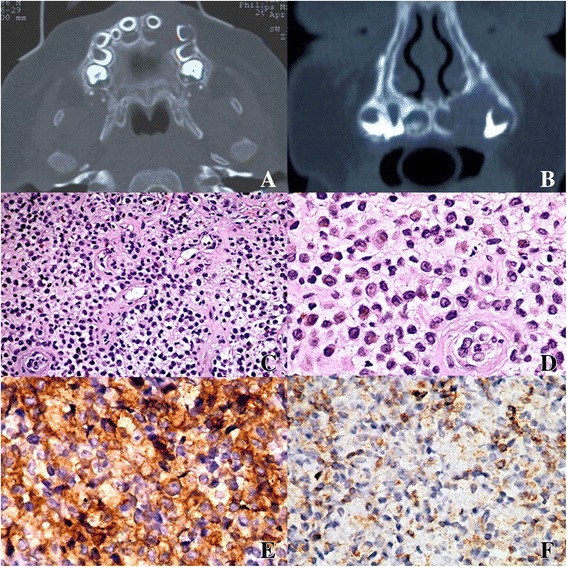
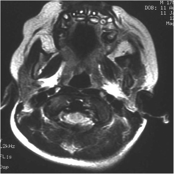
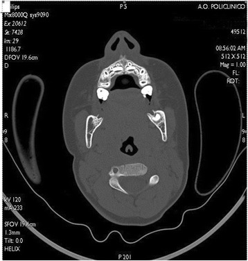
Case presentation: We report a case of Langerhans cell histiocytosis, which presented as an osteolytic lesion of the left pre-maxillae enclosing the deciduous incisor and canine in a 7-month-old white Italian boy. He was treated with chemotherapy. He achieved complete remission after 7 months and after 24 months no signs of recurrence were observed.
Conclusions: As a result of this treatment, anesthetic sequelae and loss of teeth were avoided; in addition, we prevented a loss of the vertical dimension of occlusion.
Keywords: Case report; Chemotherapy treatment; Children; Langerhans cell histiocytosis.
Figures
Similar articles
-
Langerhans' cell histiocytosis: case reports and literature review.Pediatr Dent. 1996 Jan-Feb;18(1):11-6. Pediatr Dent. 1996. PMID: 8668563 Review. No abstract available.
-
Clinical, pathological and radiological evaluation of disseminated Langerhans' cell histiocytosis in a 30-month-old boy.Dentomaxillofac Radiol. 2007 Dec;36(8):526-9. doi: 10.1259/dmfr/71449570. Dentomaxillofac Radiol. 2007. PMID: 18033951
-
Perianal presentation of Langerhans cell histiocytosis in children.Gastroenterol Clin Biol. 2010 Jan;34(1):95-7. doi: 10.1016/j.gcb.2009.06.016. Epub 2009 Oct 27. Gastroenterol Clin Biol. 2010. PMID: 19864102
-
Vulvar Lesions in an 8-Year-Old Girl: Cutaneous Manifestations of Multisystem Langerhans Cell Histiocytosis.J Pediatr Adolesc Gynecol. 2018 Apr;31(2):153-155. doi: 10.1016/j.jpag.2017.09.010. Epub 2017 Oct 6. J Pediatr Adolesc Gynecol. 2018. PMID: 28993226
-
Juvenile xanthogranuloma developing after treatment of Langerhans cell histiocytosis: case report and literature review.Int J Clin Exp Pathol. 2012;5(7):720-5. Epub 2012 Sep 5. Int J Clin Exp Pathol. 2012. PMID: 22977671 Free PMC article. Review.
Cited by
-
Efficacy of 4-year treatment of icon infiltration resin on postorthodontic white spot lesions.BMJ Case Rep. 2018 Jul 18;2018:bcr2018225639. doi: 10.1136/bcr-2018-225639. BMJ Case Rep. 2018. PMID: 30021744 Free PMC article.
-
Evaluation of Vitamin D (25OHD), Bone Alkaline Phosphatase (BALP), Serum Calcium, Serum Phosphorus, Ionized Calcium in Patients with Mandibular Third Molar Impaction. An Observational Study.Nutrients. 2021 Jun 4;13(6):1938. doi: 10.3390/nu13061938. Nutrients. 2021. PMID: 34200107 Free PMC article.
-
Revealing a rare inflammatory oral manifestation in a 6-year-old child.BMJ Case Rep. 2019 Jul 23;12(7):e229483. doi: 10.1136/bcr-2019-229483. BMJ Case Rep. 2019. PMID: 31340944 Free PMC article.
-
Three decades of progress from surgery to medical therapy for isolated neuroaxis BRAF V600E-positive Langerhans cell histiocytosis management: illustrative case.J Neurosurg Case Lessons. 2021 May 10;1(19):CASE2118. doi: 10.3171/CASE2118. eCollection 2021 May 10. J Neurosurg Case Lessons. 2021. PMID: 35854832 Free PMC article.
References
-
- PDQ Pediatric Treatment Editorial Board . PDQ Cancer Information Summaries. Bethesda: National Cancer Institute; 2002. Langerhans Cell Histiocytosis Treatment (PDQ®): Health Professional Version. - PubMed
Publication types
MeSH terms
Substances
LinkOut - more resources
Full Text Sources
Other Literature Sources
Medical
Research Materials
