Review
. 2017 May-Jun;5(3):547-563.
doi: 10.1016/j.jaip.2017.01.025.

Severe Delayed Cutaneous and Systemic Reactions to Drugs: A Global Perspective on the Science and Art of Current Practice

Affiliations
Review

Severe Delayed Cutaneous and Systemic Reactions to Drugs: A Global Perspective on the Science and Art of Current Practice

Jonathan Grant Peter et al. J Allergy Clin Immunol Pract. 2017 May-Jun.

Abstract

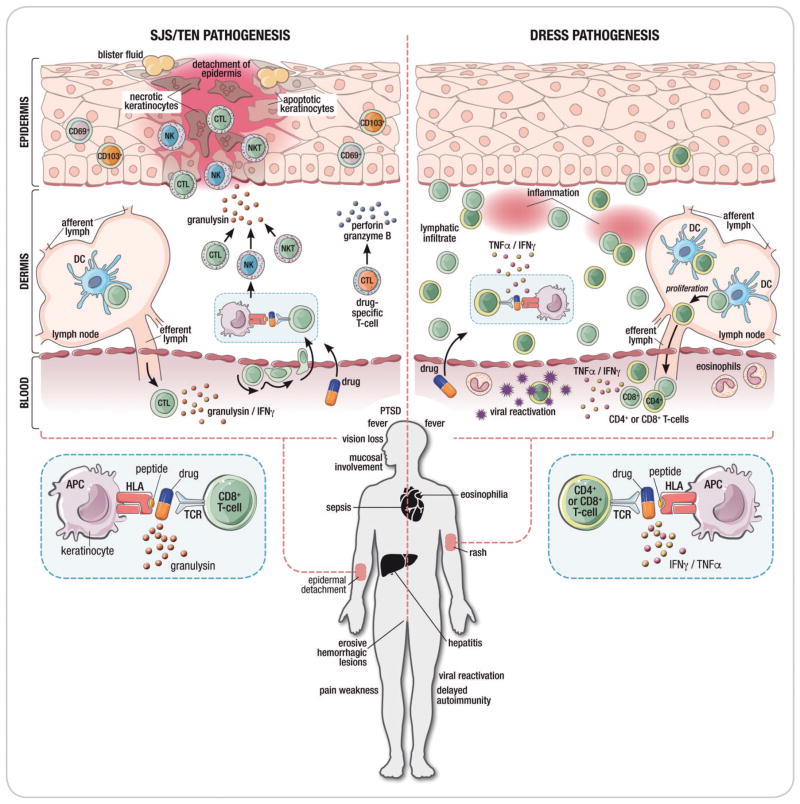
Most immune-mediated adverse drug reactions (IM-ADRs) involve the skin, and many have additional systemic features. Severe cutaneous adverse drug reactions (SCARs) are an uncommon, potentially life-threatening, and challenging subgroup of IM-ADRs with diverse clinical phenotypes, mechanisms, and offending drugs. T-cell-mediated immunopathology is central to these severe delayed reactions, but effector cells and cytokines differ by clinical phenotype. Strong HLA-gene associations have been elucidated for specific drug-SCAR IM-ADRs such as Stevens-Johnson syndrome/toxic epidermal necrolysis, although the mechanisms by which carriage of a specific HLA allele is necessary but not sufficient for the development of many IM-ADRs is still being defined. SCAR management is complicated by substantial short- and long-term morbidity/mortality and the potential need to treat ongoing comorbid disease with related medications. Multidisciplinary specialist teams at experienced units should care for patients. In the setting of SCAR, patient outcomes as well as preventive, diagnostic, treatment, and management approaches are often not generalizable, but rather context specific, driven by population HLA-genetics, the pharmacology and genetic risk factors of the implicated drug, severity of underlying comorbid disease necessitating ongoing treatments, and cost considerations. In this review, we update the basic and clinical science of SCAR diagnosis and management.

Keywords: DRESS; HLA; Immune-mediated adverse drug reactions; SJS/TEN; Severe cutaneous adverse drug reactions; T-cell.


Figures

Figure 1
Figure 1. Adverse drug reactions and the immune system
It is proposed that adverse drug reactions are classified according to their on-target vs. off-target interactions between the drug and cellular components. Both on-target and off-target effects can demonstrate concentration–exposure relationships that may differ between individuals based on acquired or genetic host factors. The interaction between the drug and the target may relate to both the dose and/or duration of treatment. On-target reactions generally relate to an augmentation of the known primary therapeutic and pharmacological action of a drug (eg. bleeding related to warfarin) and off-target effects can occur by mechanisms that are both directly immune mediated and associated with immunological memory of varied duration (drug allergy) and mechanisms without a direct immunological effect and without immunological memory that may have an “immunological phenotype”. These reactions are often mediated through a pharmacological interaction (eg. aspirin exacerbated respiratory disease or non-IgE mediated mast-cell activation seen with fluoroquinolones and opioids). Off-target reactions that are non-immunologically mediated are often dose-dependent whereas immunologically-mediated off-target reactions associated with immunological memory can be both dose dependent (T-cell mediated reactions) or dose-independent (recognition and amplification of small amounts of antigen in the case of IgE-mediated reactions). Predisposition to both on-target and off-target reactions is driven by genetic variation but also ecological factors that can vary over the course of an individual’s lifetime (Adapted from White et al (101) and Phillips (127)
Figure 2
Figure 2. The Global Epidemiology of Severe Cutaneous Adverse Drug Reactions
HLA allele frequencies are indicated for different populations and colour coded. If no prevalence figures are provided for a particular phenotype then a relevant continental dataset was not found. Abbreviations: SJS/TEN, Stevens-Johnson syndrome/toxic epidermal necrolysis; DRESS, drug reaction with eosinophilia and systemic symptoms; AGEP: Acute generalized exanthematous pustulosis; HIV SCAR: Human immunodeficiency related severe cutaneous adverse drug reactions; SSLR: Serum-sickness like reaction; NSAIDs: Non-steroidal anti-inflammatory drugs
Figure 3
Figure 3. Montage of images to illustrate important features of different clinical phenotypes
Figure 4
Figure 4. Immunopathogenesis of SCAR
Figure 4A: Proposed models of drug hypersensitivity (a) In the Hapten/prohapten model drugs form covalent bonds with endogenous proteins/peptides. These drug-modified peptides are then processed by antigen presenting cells and presented on the MHC resuting in a T-cell response. (b) The drug-specific Pharmacological-Interaction (P-I) model proposed that drug binds directly to immune receptors such as the T-cell Receptor (TCR) or HLA forming non-covalent bonds without the need for peptide. Dashed lines represent non-covalent bonds. (c) The Altered peptide repertoire model the drug forms non-covalent bonds within the binding pocket(s) of the MHC to alter the chemistry of the binding cleft and repertoire of self-peptides able to bind to the HLA molecule in question. Some of these newly presented self-peptides have not been previously tolerized and their presentation results in a T-cell response. (d) The interaction between abacavir and HLA-B*57:01 as occurs in abacavir hypersensitivity syndrome exemplifies the altered peptide repertoire model. Abacavir is shown bound to altered peptide and HLA1014 B*57:01 in the HLA-B*57:01 antigen binding cleft. Figure 4B. Proposed pathogenic mechanisms in drug-induced Stevens-Johnson syndrome/toxic epidermal necrolysis (SJS/TEN) and drug reaction with eosinophilia and systemic symptoms (DRESS). The disease process in SJS/TEN occurs in the epidermis. In SJS/TEN, the drug likely interacts with the with human leukocyte antigen protein on keratinocytes which act as antigen-presenting cells to activate drug specific CD8+ cytotoxic T cells. This interaction causes drug-specific CD8+ T cells to accumulate within epidermal blisters and release perforin and granzyme B that can kill keratinocytes. Drugs also trigger the activation of CD8+ T cells, NK cells and NKT cells to secrete granulysin which appears to be one of the most important cytotoxic mediators in SJS/TEN and can induce keratinocyte death without the need for cell contact. Clinical symptoms of SJS/TEN include a painful, blistering skin rash that results in epidermal necrosis and detachment as well as less specific symptoms including fever and sepsis. SJS/TEN is associated with a mortality of up to 50% as well as significant long-term morbidity including permanent corneal scarring, vision loss, prolonged pain and weakness, post-traumatic stress disorder and fear of drug. The dermis is the primary skin compartment involved in DRESS. Both CD8+ and CD4+ T cells are pathogenic mediators in DRESS. DRESS is characterized by a lymphatic infiltrate of T cells into the dermis and increased release of TNFα and IFNγ. DRESS is also associated with viral reactivation of human herpesviruses although the role of viral reactivation in DRESS pathogenesis is currently unclear. DRESS presents as a widespread rash of varying severity without skin separation or blistering accompanied by fever, internal organ involvement frequently affecting the liver and eosinophilia. Delayed autoimmune disease can occur as a sequela of DRESS. Abbreviations: SJS/TEN, Stevens-Johnson syndrome/toxic epidermal necrolysis; DRESS, drug reaction with eosinophilia and systemic symptoms; CTL, cytotoxic lymphocyte; NKT, natural killer T cell; NK, natural killer cell; IFNγ , interferon-gamma; TNFα, tumor necrosis factor alpha; DC, dendritic cell; APC, antigen-presenting cell; TCR, T-cell receptor; PTSD, post-traumatic stress disorder

References

    1. Kinch MS, Haynesworth A, Kinch SL, Hoyer D. An overview of FDA-approved new molecular entities: 1827–2013. Drug discovery today. 2014;19(8):1033–9. - PubMed
    1. Kongkaew C, Noyce PR, Ashcroft DM. Hospital admissions associated with adverse drug reactions: a systematic review of prospective observational studies. The Annals of pharmacotherapy. 2008;42(7):1017–25. - PubMed
    1. Smyth RMD, Gargon E, Kirkham J, Cresswell L, Golder S, Smyth R, et al. Adverse drug reactions in children - a systematic review. PloS one. 2012;7(3):e24061-e. - PMC - PubMed
    1. Mockenhaupt M. Epidemiology of cutaneous adverse drug reactions. Chemical immunology and allergy. 2012;97:1–17. - PubMed
    1. Huang HY, Luo XQ, Chan LS, Cao ZH, Sun XF, Xu JH. Cutaneous adverse drug reactions in a hospital-based Chinese population. Clinical and experimental dermatology. 2011;36(2):135–41. - PubMed