Review
. 2017 Aug 15:628:71-80.
doi: 10.1016/j.abb.2017.05.002. Epub 2017 May 5.

Applications of NMR and computational methodologies to study protein dynamics

Affiliations
Review

Applications of NMR and computational methodologies to study protein dynamics

Chitra Narayanan et al. Arch Biochem Biophys. .

Abstract

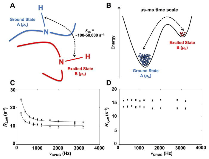
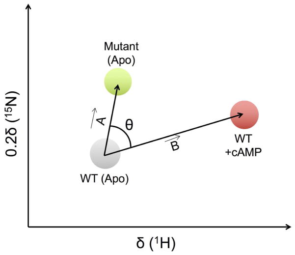
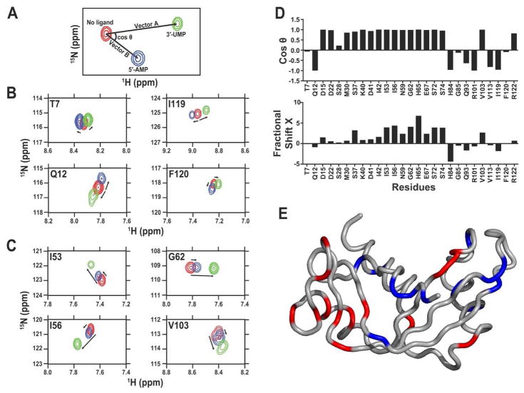
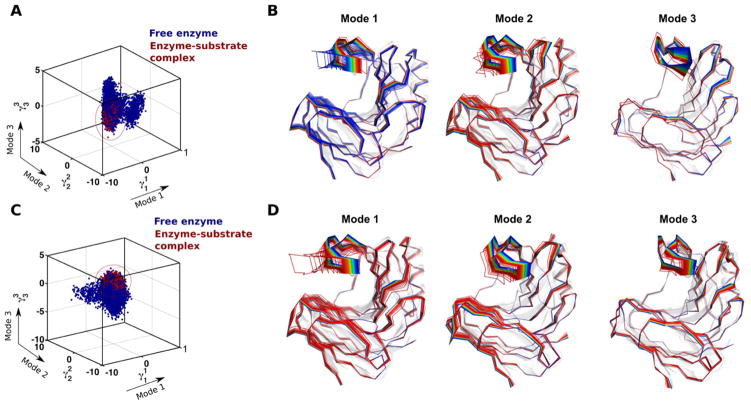
Overwhelming evidence now illustrates the defining role of atomic-scale protein flexibility in biological events such as allostery, cell signaling, and enzyme catalysis. Over the years, spin relaxation nuclear magnetic resonance (NMR) has provided significant insights on the structural motions occurring on multiple time frames over the course of a protein life span. The present review article aims to illustrate to the broader community how this technique continues to shape many areas of protein science and engineering, in addition to being an indispensable tool for studying atomic-scale motions and functional characterization. Continuing developments in underlying NMR technology alongside software and hardware developments for complementary computational approaches now enable methodologies to routinely provide spatial directionality and structural representations traditionally harder to achieve solely using NMR spectroscopy. In addition to its well-established role in structural elucidation, we present recent examples that illustrate the combined power of selective isotope labeling, relaxation dispersion experiments, chemical shift analyses, and computational approaches for the characterization of conformational sub-states in proteins and enzymes.

Keywords: Allostery; Chemical shift analysis; Conformational sub-states; Protein dynamics; Quasi anharmonic analysis; Relaxation dispersion.


Figures

Figure 1
Figure 1. Schematic depiction of exchange between two protein conformational substates on the μs-ms time scale
A) Schematic representation of a backbone residue HN vector experiencing conformational exchange between ground state A and excited state B on the μs-ms time scale. The popular Carr-Purcell-Meiboom-Gill (CPMG) and R rotating frame relaxation dispersion experiments have been collectively employed to investigate conformational exchange rates (kex) for residues experiencing conformational exchange in proteins over time frames that roughly span ~100 to ~50,000 events per second (s−1), overlapping the time scale of relevant biological events. B) Energetic representation of the two-site exchange between ground state A (higher population, pA), and excited state B (lower population pB, often invisible on fast and intermediate NMR time scales) ,,. NMR relaxation dispersion experiments can provide rates of exchange (kex), populations (pApB), and chemical shifts between interconverting species (Δω). C) Representative experimental 15N-CPMG NMR curves at 500 MHz (circles) and 800 MHz (squares) for a backbone HN vector experiencing conformational exchange on the μs-ms time scale in a protein. D) Flat experimental 15N-CPMG NMR profiles at 500 MHz (circles) and 800 MHz (squares) for a backbone HN vector that does not experience conformational exchange on the μs-ms time scale. The somewhat limited atomistic details on directionality and length-scales provided by NMR for these movements can readily be complemented by MD simulations, which offer full atomistic details of the conformational populations.
Figure 2
Figure 2. Illustration of the CHESPA approach
1H-15N resonance assignments for apo, mutant and cAMP-bound forms of EPAC are represented as grey, green, and red circles, respectively. The compounded chemical shift upon mutation and ligand binding is calculated as the magnitude of vectors A and B, respectively (see text for details). θ represents the angle between vector A and B. Figure adapted from ref. .
Figure 3
Figure 3. Application of CHESPA to human angiogenin
Projection analysis describing independent and coordinated residue variations upon 5′-AMP and 3′-UMP binding to human angiogenin subsites. (A) Graphical representation of the CHESPA approach described in ref. . The 1H-15N position of the peak is represented for the free (red) and bound forms (5′-AMP in blue and 3′-UMP in green) of the enzyme. Arrows indicate the movement of the 1H-15N chemical shift (length and direction) for each peak from its origin (in red) to its saturated position (blue and green). Projection analysis of eight selected residues responding in (B) independent or (C) coordinated manner in human angiogenin. (D) Direction cosθ and magnitude (fractional shift X) of the chemical shift perturbation of a subset of residues of angiogenin upon binding to saturation of the 5′-AMP and 3′-UMP. The cosθ quantifies the angle between vectors A (3′-UMP) and B (5′-AMP) from its initial position in the free form of the enzyme. The fractional shift X represents the fractional composite of vectors A and B induced by the ligand-induced chemical shift change. Collectiveness was observed for residues with a cosθ ~ 1, as previously described . (E) Residues showing coordinated displacements (cosθ ~ 1) are represented in red on the three-dimensional structure of human angiogenin, while residues showing uncoordinated displacements (cosθ ≠ 1) are shown in blue. Reprinted with permission from ref. . Copyright 2015 Wiley.
Figure 4
Figure 4. Conformational sub-states of xylanase B2 (XlnB2) from Streptomyces lividans determined using computational simulations and QAA
Representative conformations along the top three modes for the interconversion between free and (A–B) X6-bound and (C–D) X9-bound XlnB2 binary complexes. A total of 200,000 conformational snapshots obtained from the MD simulations were used as input for QAA to identify the top QAA-independent component vectors for characterizing the primary dynamics associated with the substrate binding process in XlnB2. Reprinted with permission from ref. . Copyright 2016 American Chemical Society.

Similar articles

Cited by

References

    1. Bhabha G, Lee J, Ekiert DC, Gam J, Wilson IA, Dyson HJ, Benkovic SJ, Wright PE. A dynamic knockout reveals that conformational fluctuations influence the chemical step of enzyme catalysis. Science. 2011;332:234–238. - PMC - PubMed
    1. Hammes-Schiffer S, Benkovic SJ. Relating protein motion to catalysis. Annu Rev Biochem. 2006;75:519–541. - PubMed
    1. Luk LYP, Javier Ruiz-Pernía J, Dawson WM, Roca M, Loveridge EJ, Glowacki DR, Harvey JN, Mulholland AJ, Tuñón I, Moliner V, Allemann RK. Unraveling the role of protein dynamics in dihydrofolate reductase catalysis. Proc Natl Acad Sci U S A. 2013;110:16344–16349. - PMC - PubMed
    1. Warner LR, Gatzeva-Topalova PZ, Doerner PA, Pardi A, Sousa MC. Flexibility in the periplasmic domain of BamA is important for function. Structure. 2016 In Press. - PMC - PubMed
    1. Palmer AG, Massi F. Characterization of the dynamics of biomacromolecules using rotating-frame spin relaxation NMR spectroscopy. Chem Rev. 2006;106:1700–1719. - PubMed

Publication types