. 2017 Jul;6(5):311-322.
doi: 10.1530/EC-17-0059. Epub 2017 May 8.

Thyroid states regulate subcellular glucose phosphorylation activity in male mice

Affiliations

Thyroid states regulate subcellular glucose phosphorylation activity in male mice

Flavia Letícia Martins Peçanha et al. Endocr Connect. 2017 Jul.

Abstract

The thyroid hormones (THs), triiodothyronine (T3) and thyroxine (T4), are very important in organism metabolism and regulate glucose utilization. Hexokinase (HK) is responsible for the first step of glycolysis, catalyzing the conversion of glucose to glucose 6-phosphate. HK has been found in different cellular compartments, and new functions have been attributed to this enzyme. The effects of hyperthyroidism on subcellular glucose phosphorylation in mouse tissues were examined. Tissues were removed, subcellular fractions were isolated from eu- and hyperthyroid (T3, 0.25 µg/g, i.p. during 21 days) mice and HK activity was assayed. Glucose phosphorylation was increased in the particulate fraction in soleus (312.4% ± 67.1, n = 10), gastrocnemius (369.2% ± 112.4, n = 10) and heart (142.2% ± 13.6, n = 10) muscle in the hyperthyroid group compared to the control group. Hexokinase activity was not affected in brain or liver. No relevant changes were observed in HK activity in the soluble fraction for all tissues investigated. Acute T3 administration (single dose of T3, 1.25 µg/g, i.p.) did not modulate HK activity. Interestingly, HK mRNA levels remained unchanged and HK bound to mitochondria was increased by T3 treatment, suggesting a posttranscriptional mechanism. Analysis of the AKT pathway showed a 2.5-fold increase in AKT and GSK3B phosphorylation in the gastrocnemius muscle in the hyperthyroid group compared to the euthyroid group. Taken together, we show for the first time that THs modulate HK activity specifically in particulate fractions and that this action seems to be under the control of the AKT and GSK3B pathways.

Keywords: T3 action; glucose metabolism; mitochondria; muscle; thyroid.


Figures

Figure 1
Figure 1
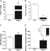
Assessment of hormonal status and markers of hyperthyroidism in mice. Hyperthyroidism was induced by treating animals with T3, as described in the ‘Materials and methods’ section. (A) Total T3 and (B) total T4 levels were measured using RIA. (C) HW/BW (heart weight/body weight) was obtained by dividing heart weight (mg) by body weight (g). (D) Liver D1 mRNA expression was assayed by real-time PCR and normalized to the housekeeping gene 36β4. (Inset) Ct values from housekeeping gene 36β4. Values are means ± s.e.m. of 3–7 animals per experimental group. Open bars represent control, and black bars, T3-administered groups. #P < 0.05, ***P < 0.0001 compared to the euthyroid group (Eu).
Figure 2
Figure 2
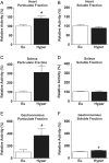
Subcellular HK activity in mouse tissues. Hyperthyroidism was induced by treating animals with T3 for 21 days, and subcellular fractions were prepared from heart (A, B) soleus (C, D) and gastrocnemius (E, F) muscle. HK activity was assessed as described in the ‘Materials and methods’ section in the particulate (A, C and E) and soluble (B, D, F) fractions. Values are means ± s.e.m. of 10 animals per experimental group. Open bars represent euthyroid, and black bars, hyperthyroid groups. #P < 0.05, *P < 0.01 compared to the euthyroid group (Eu).
Figure 3
Figure 3
Effect of TH treatment on HK activity from C2C12 myotubes. C2C12 cells were differentiated as described in the ‘Materials and methods’ section for 7 days. The cells were treated with vehicle (open bars) or 1.85 nM T3 (black bars). HK activity was assessed as described in the ‘Materials and methods’ section in the soluble (A) and particulate (B) fractions. Values are means ± s.e.m. of 3 independent experiments. #P < 0.05 compared to the vehicle (control).
Figure 4
Figure 4
Acute T3 treatment on subcellular HK activity in gastrocnemius muscle. Mice were treated with a single T3 dose (1.25 µg/g) and killed at the indicated time points after T3 injection. HK activity was assessed as described in the ‘Materials and methods’ section in the particulate (A) and soluble (B) fractions. Values are means ± s.e.m. of 4 animals for each time point and group. Open bars represent control, and black bars, T3-administered groups. #P < 0.05 compared to the euthyroid group.
Figure 5
Figure 5
HK2 mRNA and protein levels in mouse gastrocnemius muscle. Hyperthyroidism was induced by treating animals with T3 for 21 days. Hexokinase II mRNA (A) and protein (B) levels were performed using real-time PCR and Western blot, respectively, as described in ‘Materials and methods’ section. Values are means ± s.e.m. of 3–5 animals per experimental group. Open bars represent euthyroid, and black bars, hyperthyroid groups. ***P < 0.0001 compared to the euthyroid group.
Figure 6
Figure 6
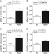
T3 induced AKT and GSK3B in mouse gastrocnemius muscle. Hyperthyroidism was induced by treating animals with T3 for 21 days. Western blots were performed as described in the ‘Materials and methods’ section. (A) Phospho AKT, (B) total AKT, (C) phospho GSK3B and (D) total GSK3B protein levels. Values are means ± s.e.m. of 3–5 animals per experimental group. Open bars represent euthyroid, and black bars, hyperthyroid groups. *P < 0.01 compared to the euthyroid group.
Figure 7
Figure 7
Mechanism of thyroid hormone action on hexokinase activity. Thyroid hormones can exert their effects through the nuclear thyroid hormone receptor, cytoplasmic thyroid hormone receptor or membrane hormone receptor (integrin αvβ3). Both nuclear and cytoplasmic TH receptors are able to participate in AKT phosphorylation. Once activated, Akt phosphorylates HK, increasing its association with mitochondria. In parallel to this effect, Akt also phosphorylates and inactivates GSK3B. This inactivation may decrease VDAC phosphorylation, allowing more HK binding to mitochondria. Taken together, these effects result in increased mitochondrial hexokinase activity. It seems that the ERK pathway does not have a relevant effect on HK activity.

Similar articles

Cited by

References

    1. Bianco AC, Maia AL, da Silva WS, Christoffolete MA. Adaptive activation of thyroid hormone and energy expenditure. Bioscience Reports 2005. 25 191–208. (10.1007/s10540-005-2885-6) - DOI - PubMed
    1. Sugden MC, Liu YL, Holness MJ. Glucose utilization by skeletal muscles in vivo in experimental hyperthyroidism in the rat. Biochemical Journal 1990. 271 421–425. (10.1042/bj2710421) - DOI - PMC - PubMed
    1. Harper M, Ballantyne S, Leach M, Brand MD. Effects of thyroid hormones on oxidative phosphorylation. Biochemical Journal 1993. 21 785–792. (10.1042/bsbib210785) - DOI - PubMed
    1. Harper ME, Brand MD. Hyperthyroidism stimulates mitochondrial proton leak and ATP turnover in rat hepatocytes but does not change the overall kinetics of substrate oxidation reactions. Canadian Journal of Physiology and Pharmacology 1994. 72 899–908. (10.1139/y94-127) - DOI - PubMed
    1. Gordon A, Swartz H, Shwartz H. 3,5,3′ Triiodo-L-thyronine stimulates 2-deoxy-D-glucose transport into L6 muscle cells through the phosphorylation of insulin receptor beta and the activation of PI-3k. Thyroid 2006. 16 521–529. (10.1089/thy.2006.16.521) - DOI - PubMed

LinkOut - more resources